

Bleeding canker on red oak (Quercus rubra)
Photo Gallery
Site will be retired 9/1/2026
This site is no longer being developed and will be retired on September 1, 2026. Please contact us if you have any questions or would like to provide support to continue the project.
|
P. cactorum bleeding canker 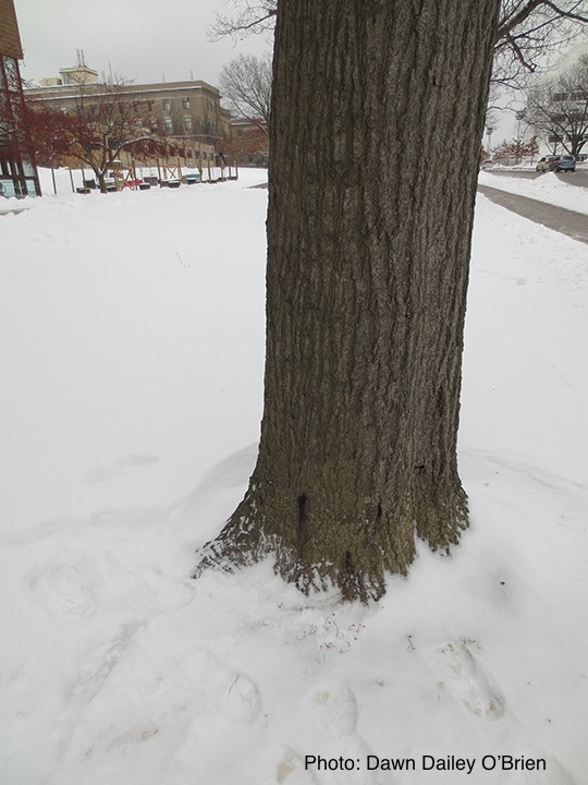 |
P. tentaculata hyphae 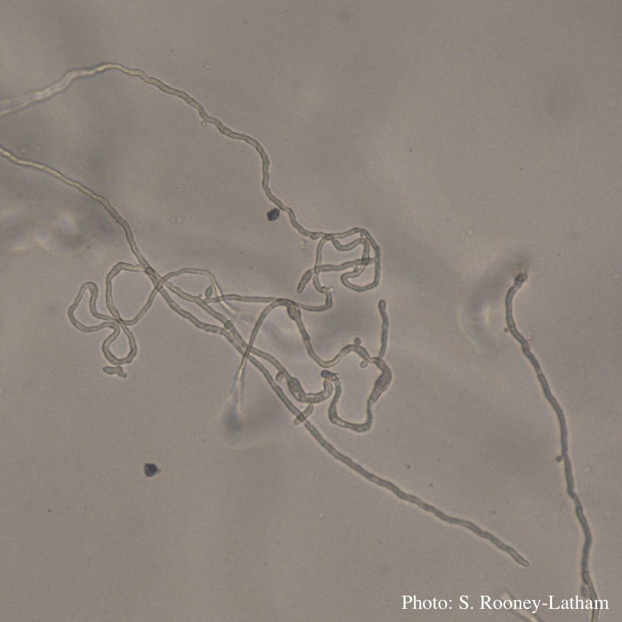 Looping hyphae commonly seen with P. tentaculata on PARP media |
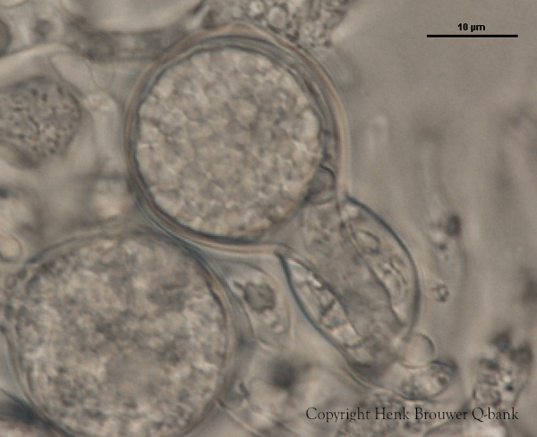
P. ramorum oogonium 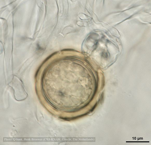 Oogonium with thick oogonial wall, photo from Q-bank, used with permission |
|
P. alni canker on gray alder 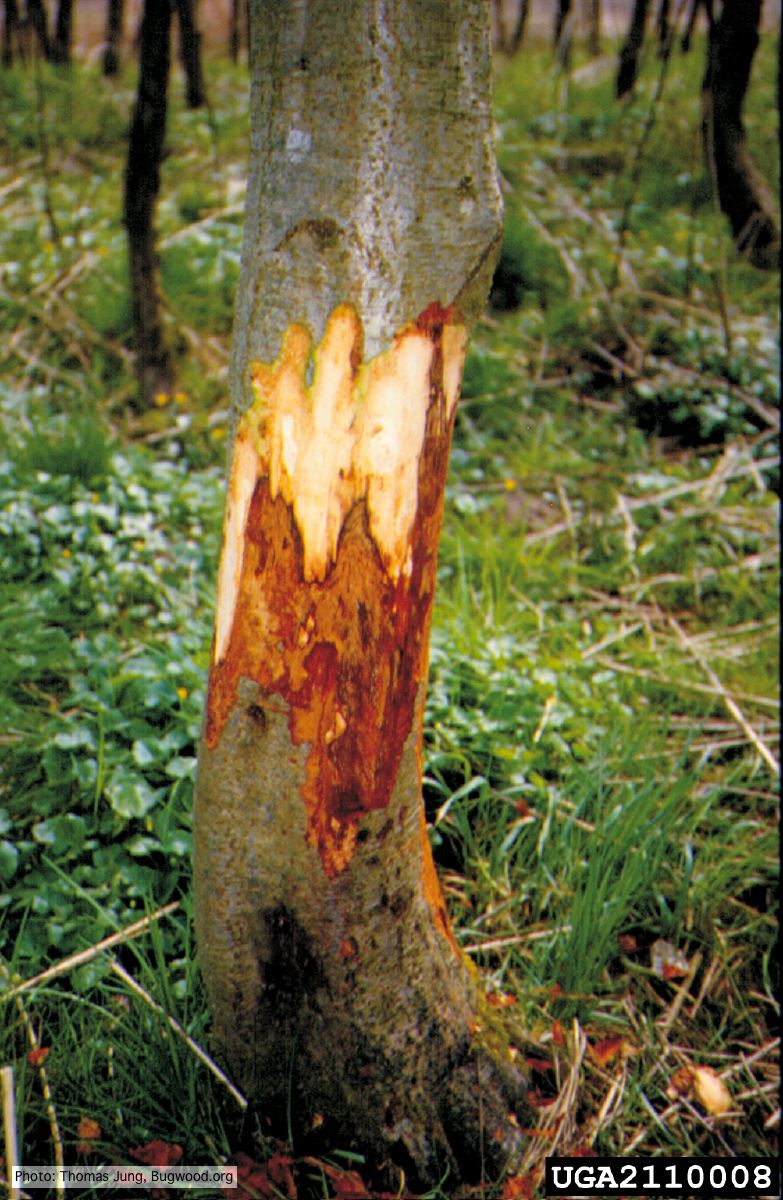 Grey alder (A. incana) with collar rot caused by P. alni |
P. tentaculata disease symptoms on sticky monkey flower 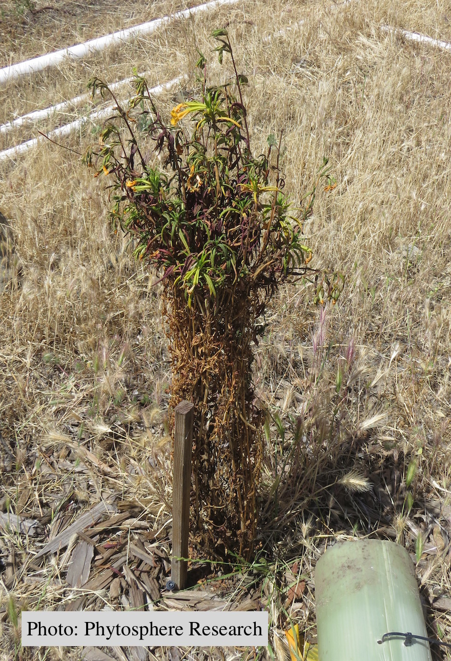 Outplanted sticky monkey flower (Diplacus aurantiacus) infected with P. |
P. nicotianae oogonia 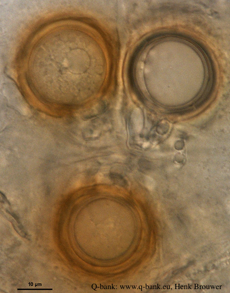 P. nicotianae oogonia 100x. Photo from Q-bank: www.q-bank.eu, Henk Brouwer (CBS-KNAW, Utrecht, The Netherlands) |
|
P. cambivora on dead and dying chinquapin  Dead and dying chinquapin infected with P. cambivora |
P. pseudosyringae hyphal swellings  Sub-globose hyphal swellings in water |
P. megakarya oospore
P. megakarya oogonia, oospore, and antheridium
|
|
Basal canker on Port Orford Cedar stump  |
P. kernoviae leaf wilt 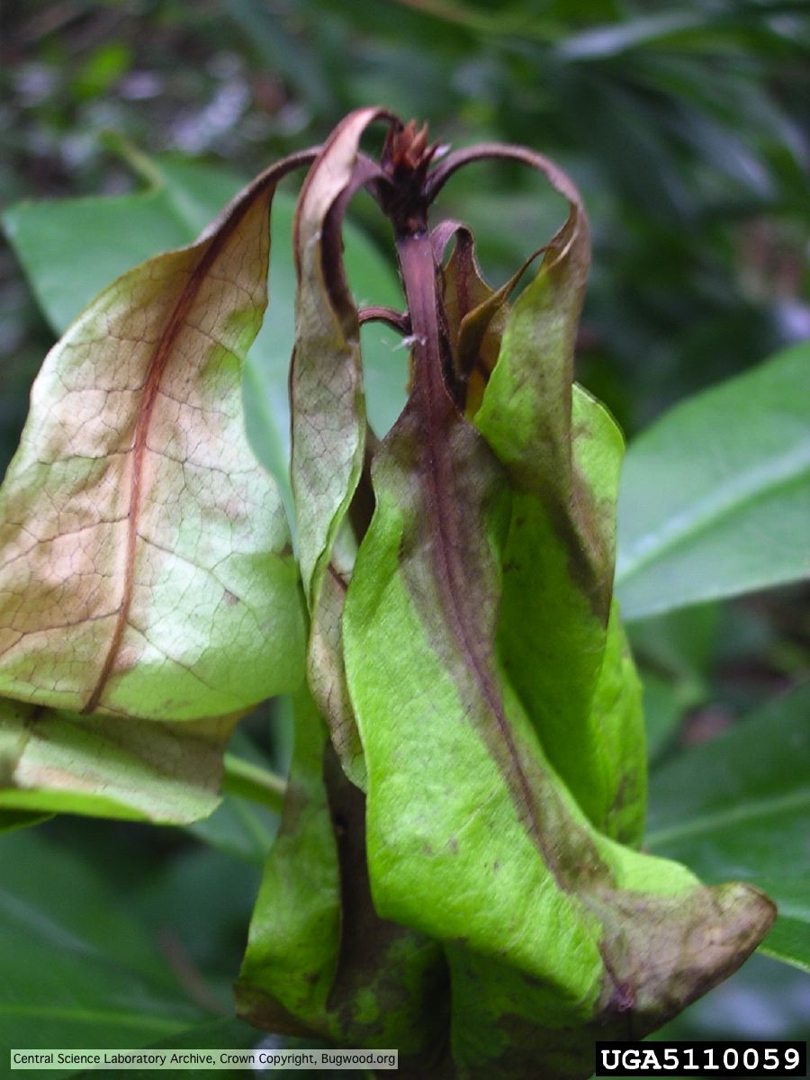 Necrosis of rhododendron leaves. |
Phytophthora cactorum canker on beech tree  Canker on Fagus sp. caused by Phytophthora cactorum. |