

P. ramorum sporangium
Photo Gallery
Site will be retired 9/1/2026
This site is no longer being developed and will be retired on September 1, 2026. Please contact us if you have any questions or would like to provide support to continue the project.
|
P. ramorum sporangium 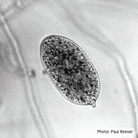 |
Mat to control spread of P. agathidicida  Jogger running over a plastic-reinforced, foam-mat containing a 2% solution of Trigene™ Advance (quaternary ammonium compound) as part of a cross-country event, in the Waitakere Regional Park |
P. agathidicia sporangia 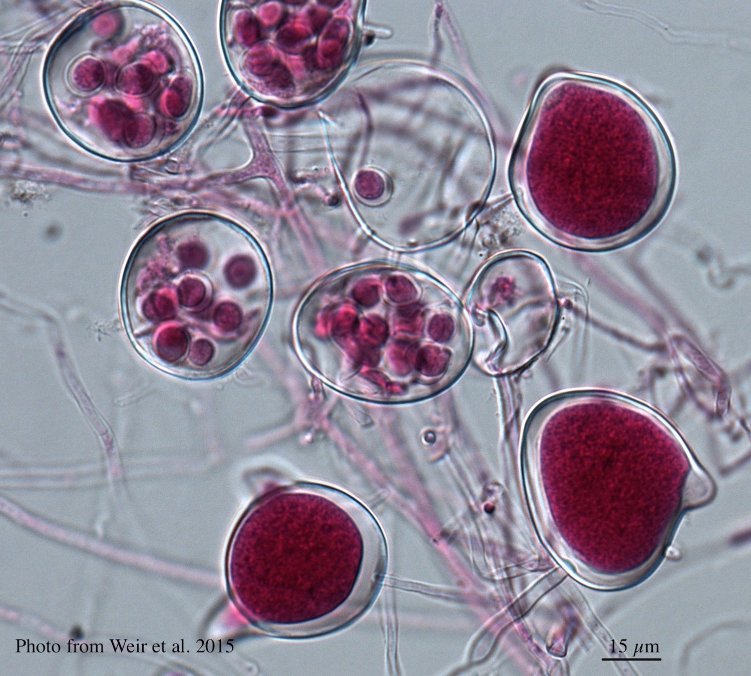 Differentiation of the cytoplasm within papillate sporangia into acid fuchsin stained zoospores |
|
P. pluvialis symptoms 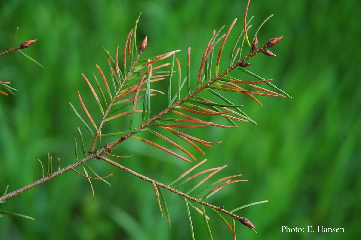 Symptoms of red needle cast on Douglas-fir needles |
P. pinifolia on Pinus radiata 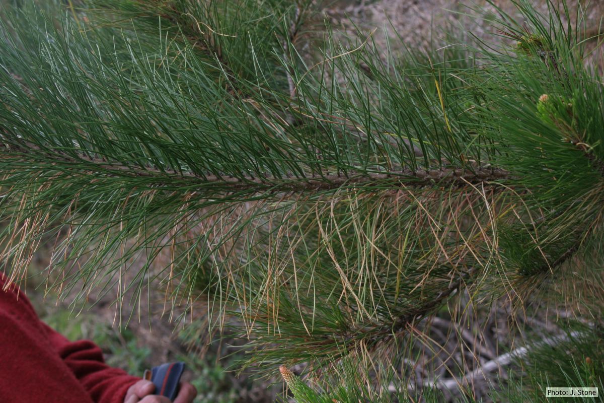 Dead needles on lower side of P. radiata branch. |
P. tentaculata disease symptoms on sticky monkey flower 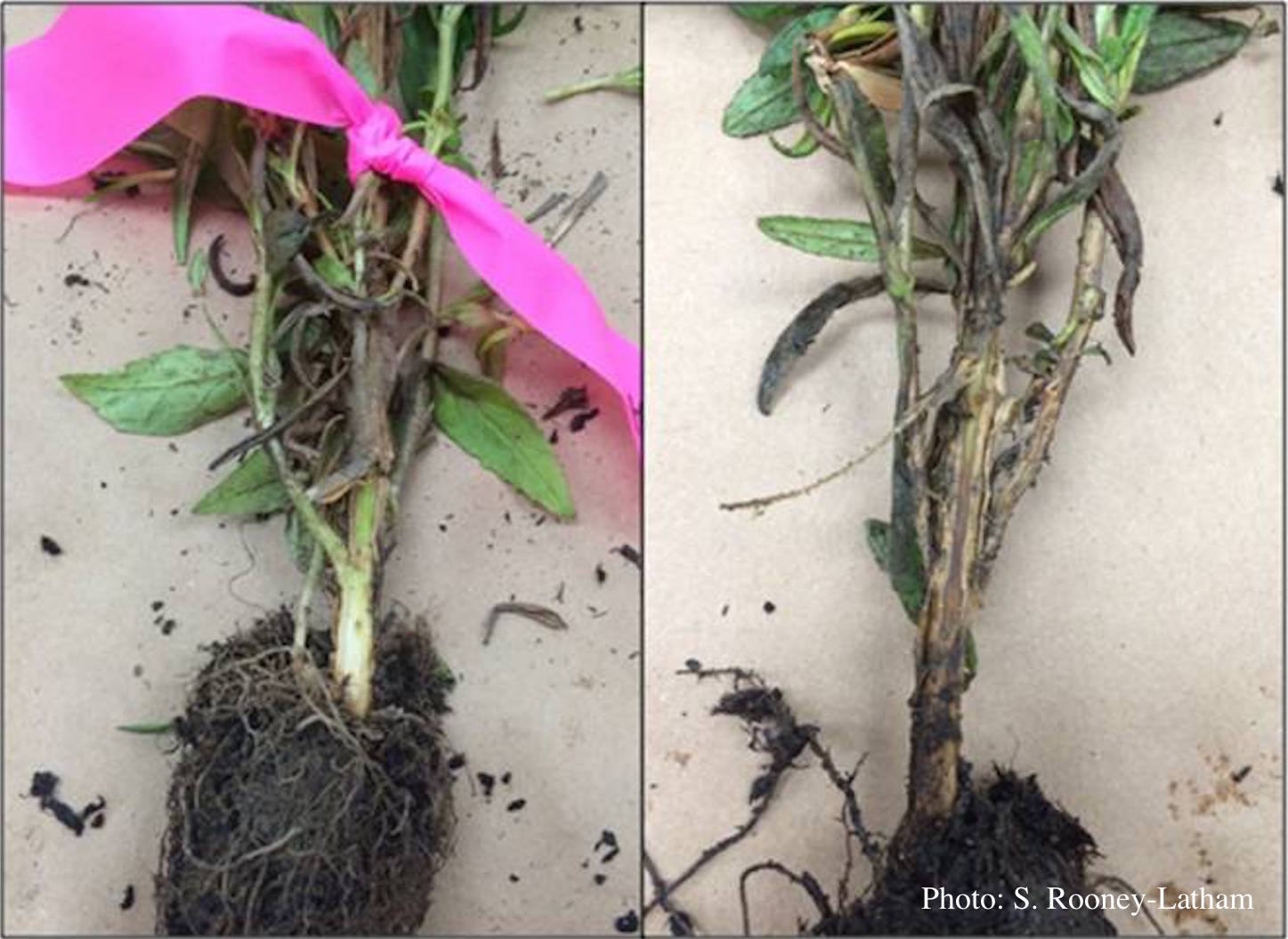 Crown and root rot (left) on sticky monkey flower (Diplacus aurantiacus) compared with a control (right) |
|
P. palmivora oogonia  P. palmivora oogonia with antheridia |
P. pluvialis on Pinus radiata in New Zealand 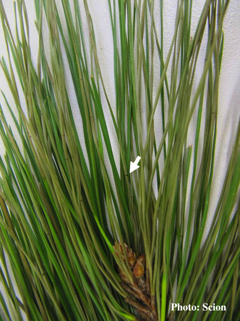 A Pinus radiata needle showing faded olive- or khaki- coloured lesions consistent with the presence of red needle cast disease. Arrow shows resinous bands within the extended olive lesion. |
P. cactorum colony morphology on V8  Colony morphology on V8 at 14 days |
|
P. tentaculata sporangium 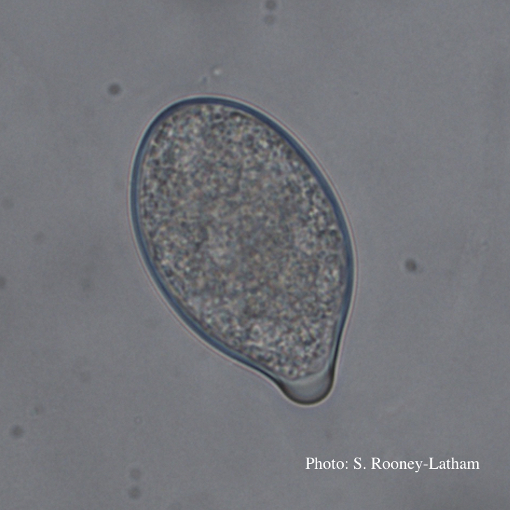 Papillate sporangium of P. tentaculata |
Phytophthora taxon Agathis bole canker  Canker on a Kauri tree, New Zealand |
P. austrocedrae - Mal del ciprés, stages of decline  Colony morphology of P. austrocedrae at 16 C after four weeks on PDA |