

Fig. 40. P. alni subsp. uniformis. Fig. 41. P. alni subsp. multiformis German variant. Fig. 42. P. alni subsp. alni. Fig. 43.
Photo Gallery
Site will be retired 9/1/2026
This site is no longer being developed and will be retired on September 1, 2026. Please contact us if you have any questions or would like to provide support to continue the project.
|
P. alni oogonia subspecies and variants 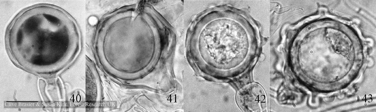 |
P. chlamydospora chlamydospore 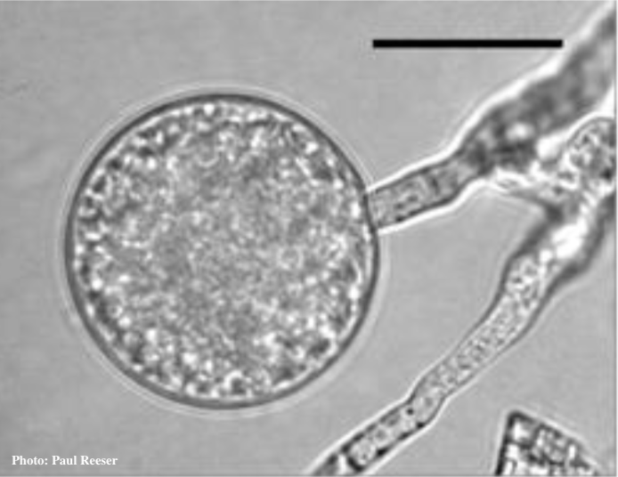 Phytophthora chlamydospora chlamydospore in agar. Bar is 20 µm. |
P. austrocedrae - sporangia drawings 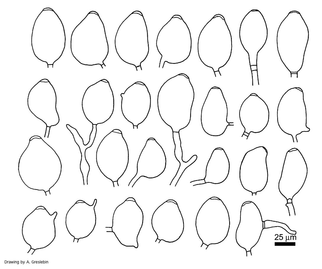 Phytophthora austrocedrae. Morphology of sporangia. Bar: 25 mm. Greslebin et al. 2007 |
|
P. cambivora colony morphology on MA 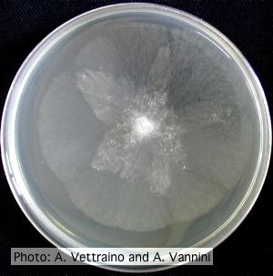 Appressed stellate colony morphology at 14 days at 20°C on MA |
P. pinifolia colony morphology on PDA 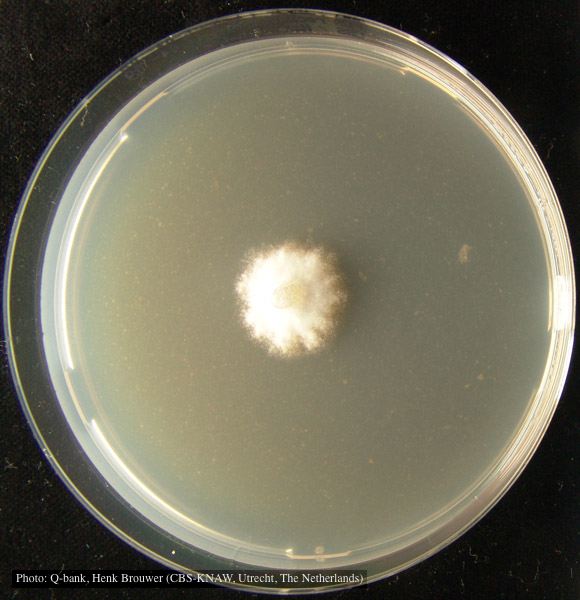 Colony pattern after 7 days on PDA at 24C, photo from Q-bank, used with permission. |
P. pluvialis symptoms on Douglas-fir 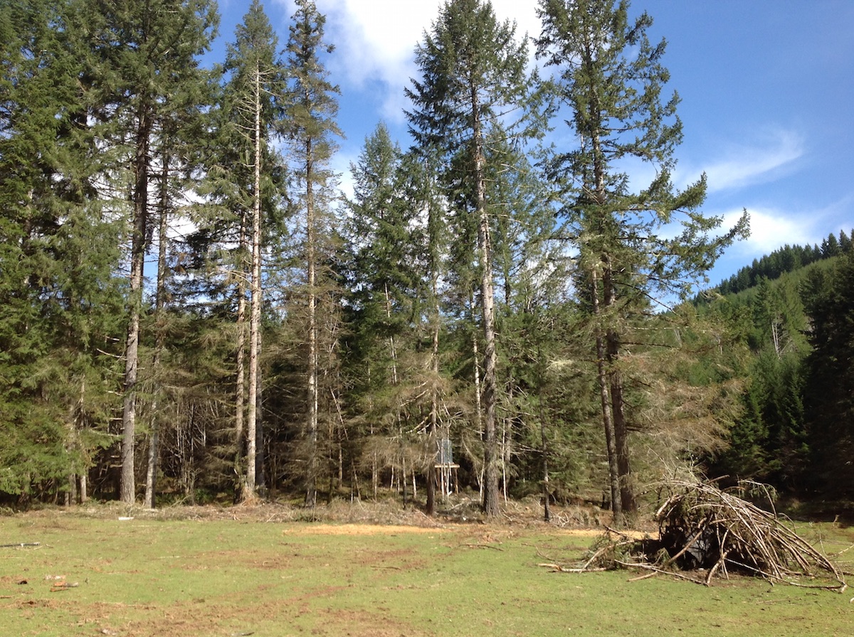 P. pluvialis symptoms of red needle cast on Douglas-fir, western Oregon 2015 |
|
Necrotic lesion in phloem caused by P. austrocedrae  Necrotic lesion in phloem with resin pocket caused by P. austrocedrae |
P. ramorum leaf symptoms on tan oak 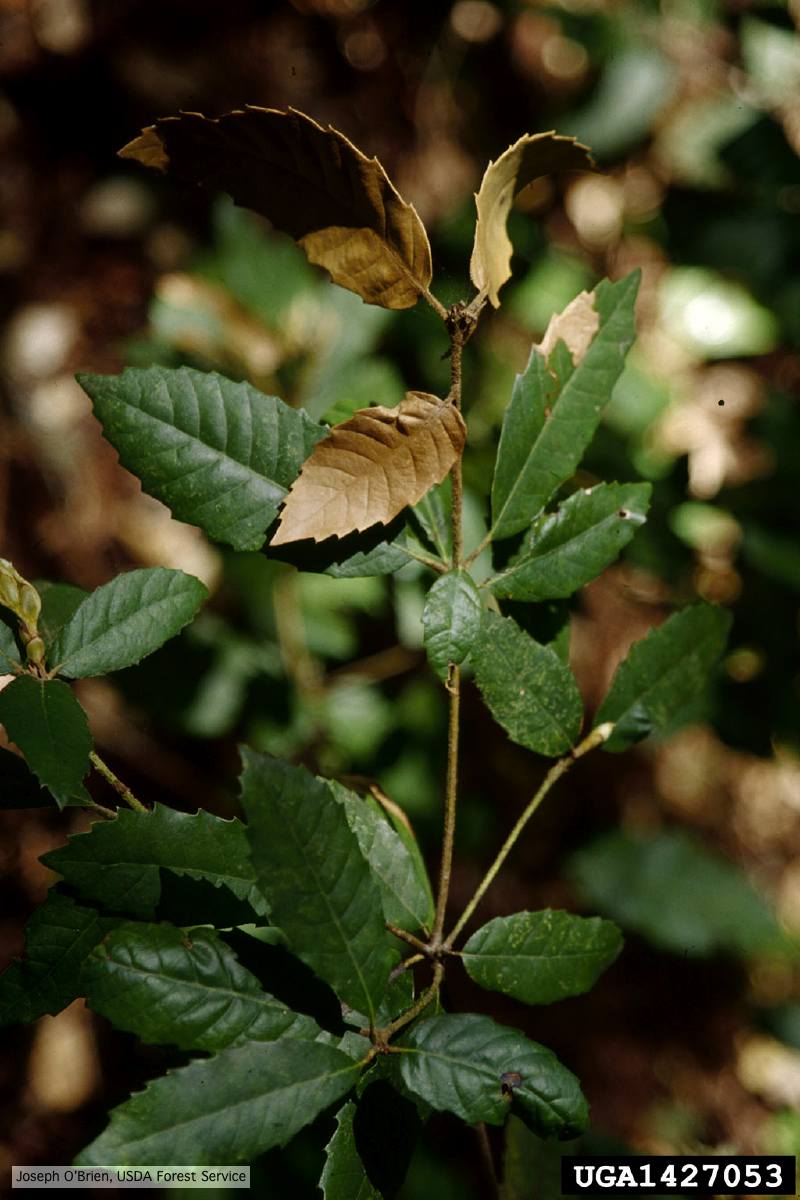 Tip symptoms on tanoak seedling (Notholithocarpus densiflorus). |
P. pinifolia on Pinus radiata 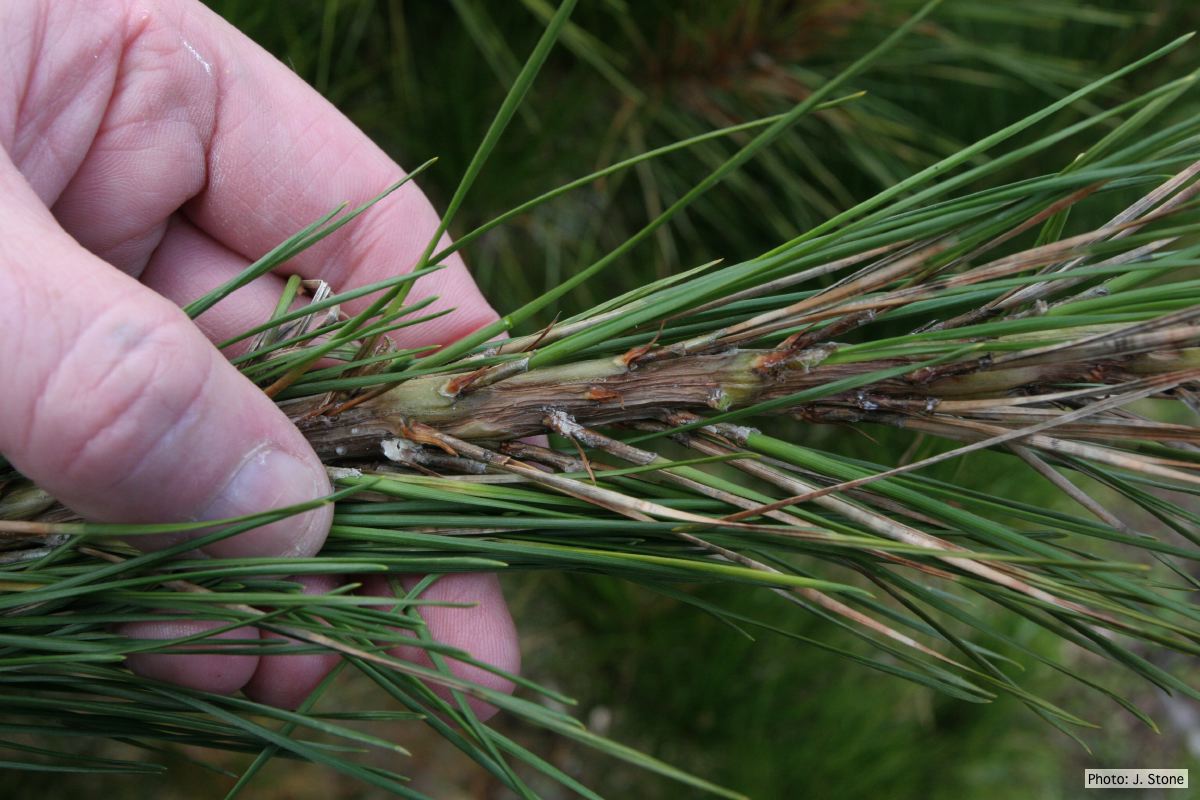 Pinus radiata needles, note “black line” symptom near needle bases |
|
Growth of P. megakarya on PDA 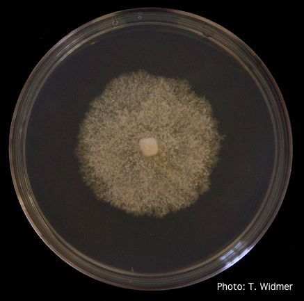 Growth of P. megakarya on potato dextrose agar |
P. lateralis colony morphology on PDA 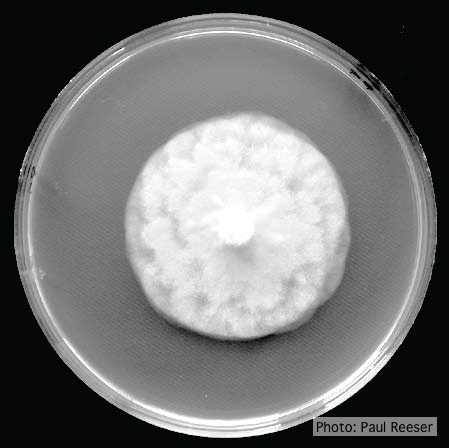 Growth morphology on PDA of Phytophthora lateralis |
P. tentaculata chlamydospore 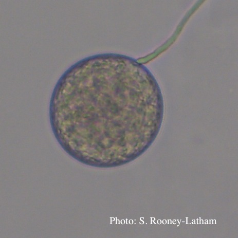 Terminal chlamydospore of P. tentaculata |