

Colony morphology on V8 at 14 days
Photo Gallery
Site will be retired 9/1/2026
This site is no longer being developed and will be retired on September 1, 2026. Please contact us if you have any questions or would like to provide support to continue the project.
|
P. cactorum colony morphology on V8  |
P. siskiyouensis bleeding canker  Bole lesions in the tissues under the bark of a bleeding canker: discoloration in the secondary phloem tissue |
Dead Port Orford Cedar 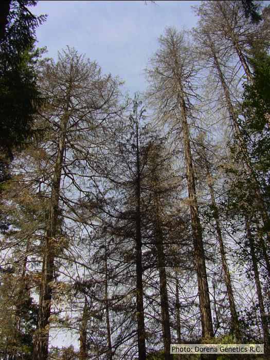 Dead Chamaecyparis lawsoniana, BLM Roseburg District in Oregon |
|
P. ramorum Dieback and shoot blight on Grand fir 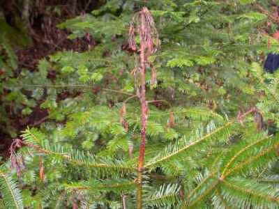 Dieback and shoot blight symptoms caused by P. ramorum on Abies grandis |
P. cambivora colony morphology on PDA 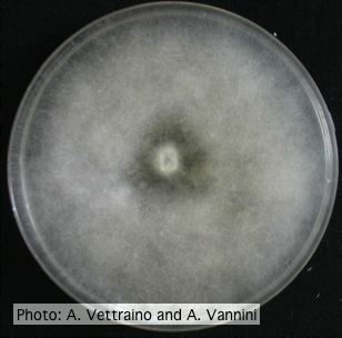 Uniform fluffy colony morphology at 14 days at 20°C on PDA |
P. cinnamomi hyphal swellings 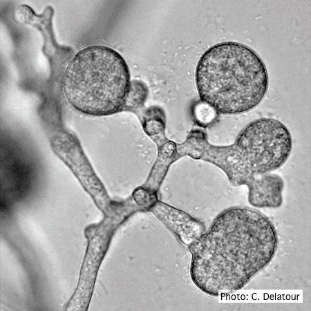 P. cinnamomi hyphal swellings (or thin walled chlamydospores) |
|
P. pinifolia on Pinus radiata  Pinus radiata, note Infected needles at right angles to stem |
P. chlamydospora sporangium  Phytophthora chlamydospora sporangium in water. Bar is 20µm. |
P. cinnamomi on Banksia 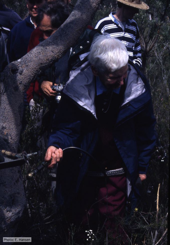 Gretna Weste injecting Banksia with phosphonate |
|
P. pinifolia colony morphology on V8 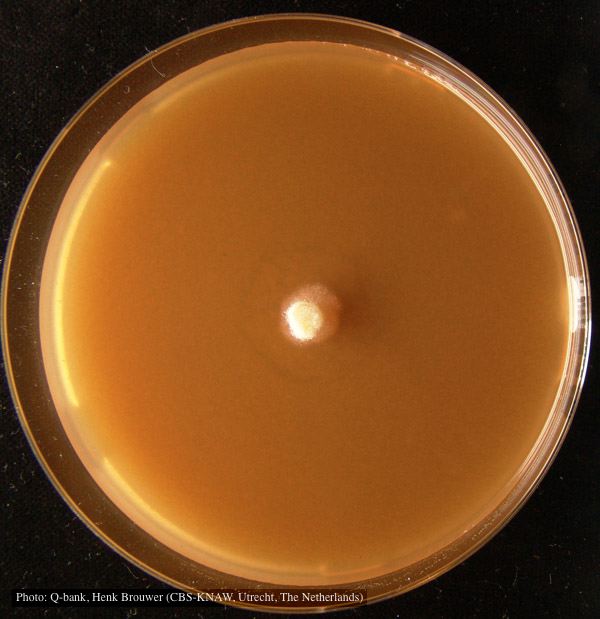 Colony pattern after 7 days on V8 at 24C, photo from Q-bank, used with permission |
P. alni symptoms on European Alder  Mature, riparian common alder (A. glutinosa) stand heavily impacted by root and collar rot caused by P. alni |
P. palmivora chlamydospore 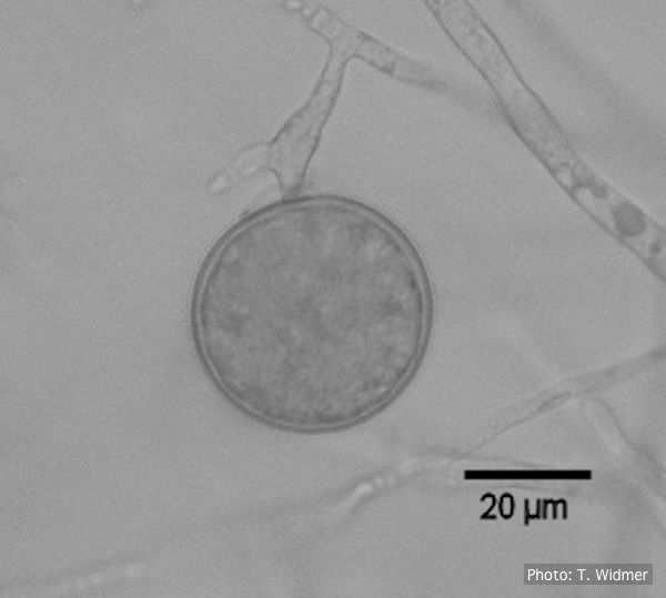 Terminal chlamydospore of P. palmivora |