

Broadly ovoid, papillate sporangia in water
Photo Gallery
Site will be retired 9/1/2026
This site is no longer being developed and will be retired on September 1, 2026. Please contact us if you have any questions or would like to provide support to continue the project.
|
P. pseudotsugae sporangia  |
Mal del ciprés, dead and dying trees along river 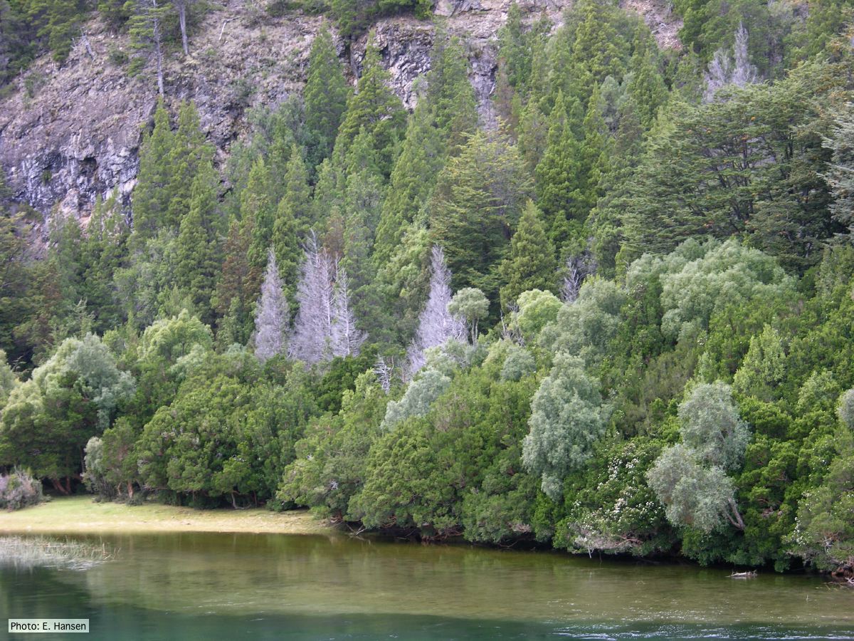 Mal del ciprés, dead and dying trees along river |
P. alni lesion in alder, Illwald, France 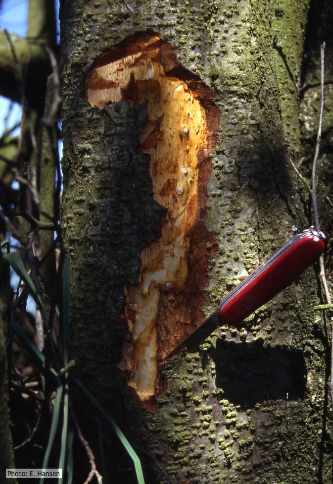 P. alni lesion in alder, Illwald, France |
|
P. alni canker on gray alder 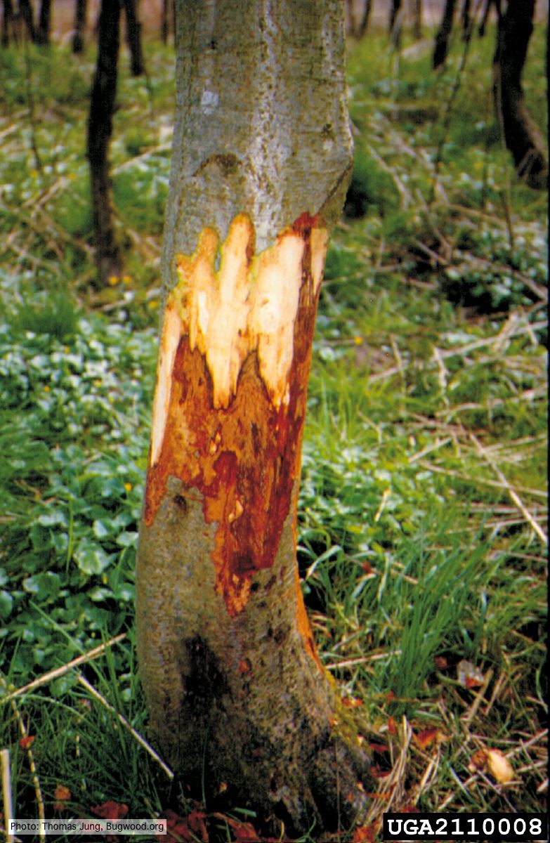 Grey alder (A. incana) with collar rot caused by P. alni |
P. megasperma oogonium 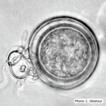 Oogonium with paragynous antheridia applied close to the ogonial stalk. |
P. siskiyouensis sporangia 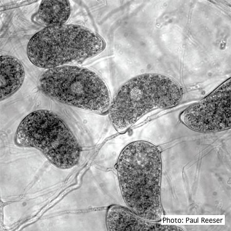 Sporangia showing a variety of shapes and orientations of semi-papillae and sporangiophores |
|
P. siskiyouensis canker on Italian alder  Bleeding canker at the base of a tree and a sprinkler emitter (arrow) adjacent to the trunk |
P. siskiyouensis oogonium with amphigynous antheridium 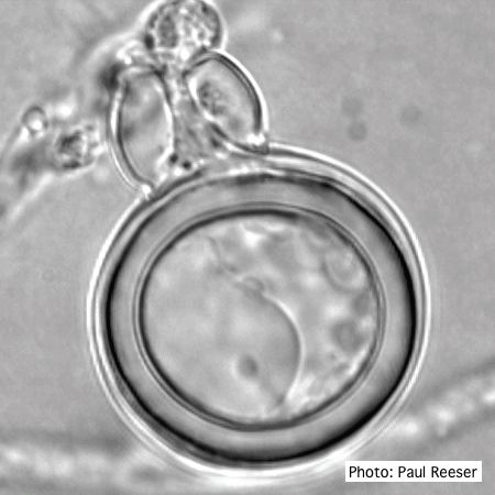 P. siskiyouensis oogonium with amphigynous antheridium |
P. frigida chlamydospore 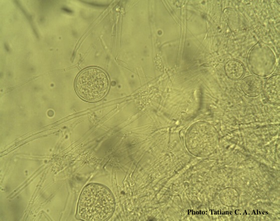 Globose chlamydospores of P. frigida |
|
P. nemorosa colony morphology on V8 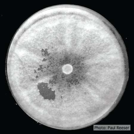 Colony morphology on V8 at 14 days |
Basal canker on Port-Orford cedar 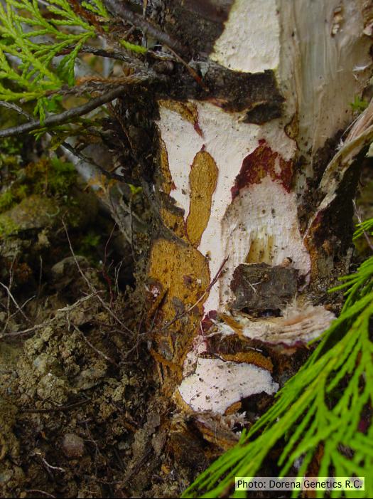 Basal canker on Chamaecyparis lawsoniana |
Port Orford cedar seedlings 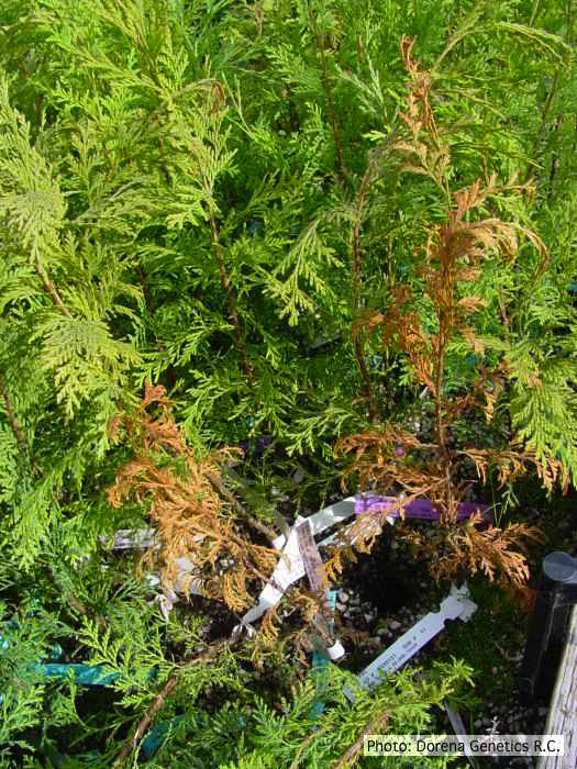 Raised beds for testing disease resistance of Port-Orford-cedar seedlings at the Dorena Genetic Resource Center |