

Ovoid, semi-papillate sporangium showing medium length pedicel.
Photo Gallery
Site will be retired 9/1/2026
This site is no longer being developed and will be retired on September 1, 2026. Please contact us if you have any questions or would like to provide support to continue the project.
|
P. nemorosa sporangium  |
P. lateralis on Port Orford cedar  Lesion caused by aerial infection on Chaemacyparis lawsoniana in Lopérec, France |
P. ramorum Dieback and shoot blight on Grand fir 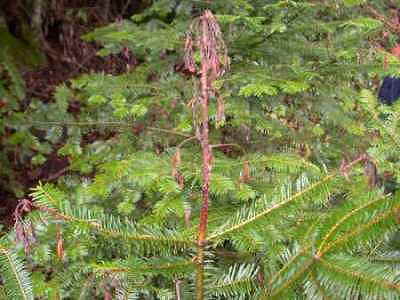 Dieback and shoot blight symptoms caused by P. ramorum on Abies grandis |
|
P. lateralis on Port Orford cedar 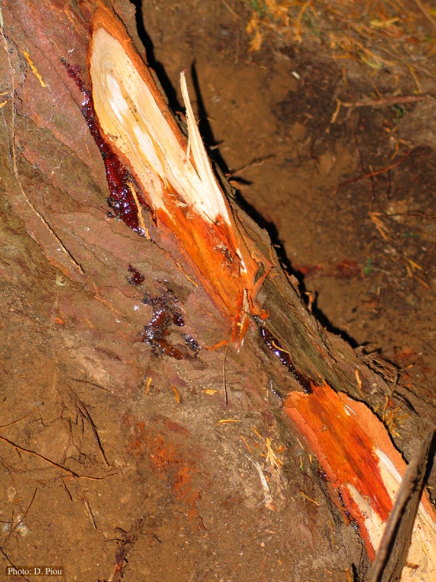 Small root lesions on Chaemacyparis lawsoniana |
P. pinifolia on Pinus radiata  Pinus radiata, note Stem canker associated with necrotic needles. |
P. tentaculata chlamydospore  P. tentaculata chlamydospore with short hyphal projection |
|
Basal canker on Port-Orford cedar 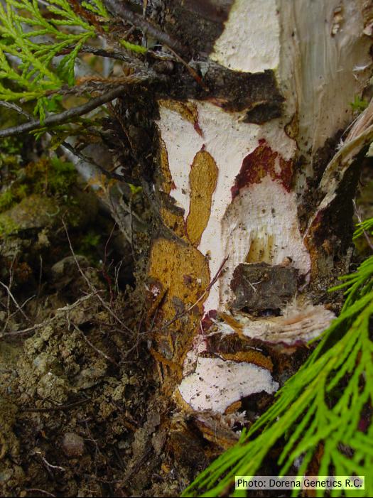 Basal canker on Chamaecyparis lawsoniana |
P. megakarya chlamydospore 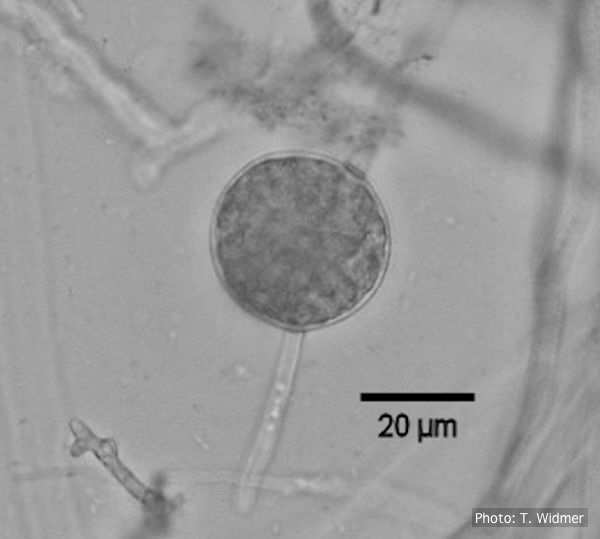
Terminal chlamydospore of P. megakarya
|
P. cambivora sporangium with internal extended proliferation  Empty sporagia showing internal nested and extended proliferation |
|
P. cambivora disease symptoms 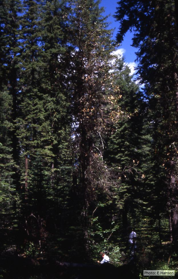 Dead and dying chinquapin infected with P. cambivora |
P. cambivora on dead and dying chinquapin  Dead and dying chinquapin infected with P. cambivora |
P. cactorum sporangia 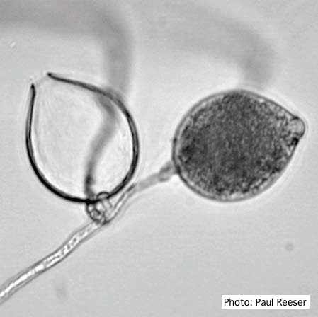 Broadly ovoid, papillate sporangia in water. |