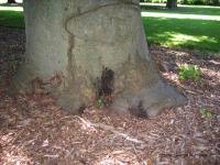
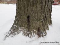
Bleeding canker on European beech (Fagus sylvatica)
Photo Gallery
|
P. cactorum bleeding canker |
P. pseudosyringae oogonium Oogonium with paragynous antheridium in agar |
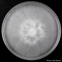
P. chlamydospora colony morphology on V8 agar P. chlamydospora colony morphology on V8 agar |
|
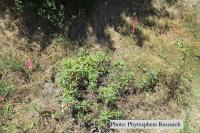
P. tentaculata disease symptoms on California mugwort Outplanted California mugwort (Artemisia douglasiana) infected with P. tentaculata, 4.5 years after planting. Plant shows stunting and chlorosis. (P. cryptogea and P. lacustris were also baited from roots/soil of this plant). |
P. siskiyouensis oogonium with paragynous antheridium P. siskiyouensis oogonium with paragynous antheridium |
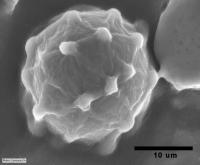
P. katsurae oogonium Micrograph of warty oogonium |
|
P. cactorum bleeding canker Bleeding canker on red oak (Quercus rubra) |
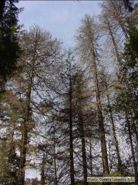
Dead Port Orford Cedar Dead Chamaecyparis lawsoniana, BLM Roseburg District in Oregon |
Growth morphology on V8 of P. lateralis Colony morphology on V8 at 14 days |
|
Phytophthora taxon Agathis bole canker Canker on a Kauri tree, New Zealand |
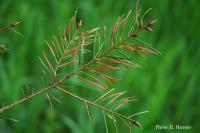
P. pluvialis symptoms Symptoms of red needle cast on Douglas-fir needles |
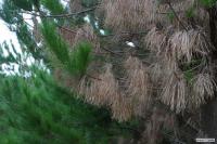
P. pinifolia on Pinus radiata Pinus radiata dead needles caused by DFP with healthy new growth from DFP |


