

Crown and root rot (left) on sticky monkey flower (Diplacus aurantiacus) compared with a control (right)
Photo Gallery
Site will be retired 9/1/2026
This site is no longer being developed and will be retired on September 1, 2026. Please contact us if you have any questions or would like to provide support to continue the project.
|
P. tentaculata disease symptoms on sticky monkey flower 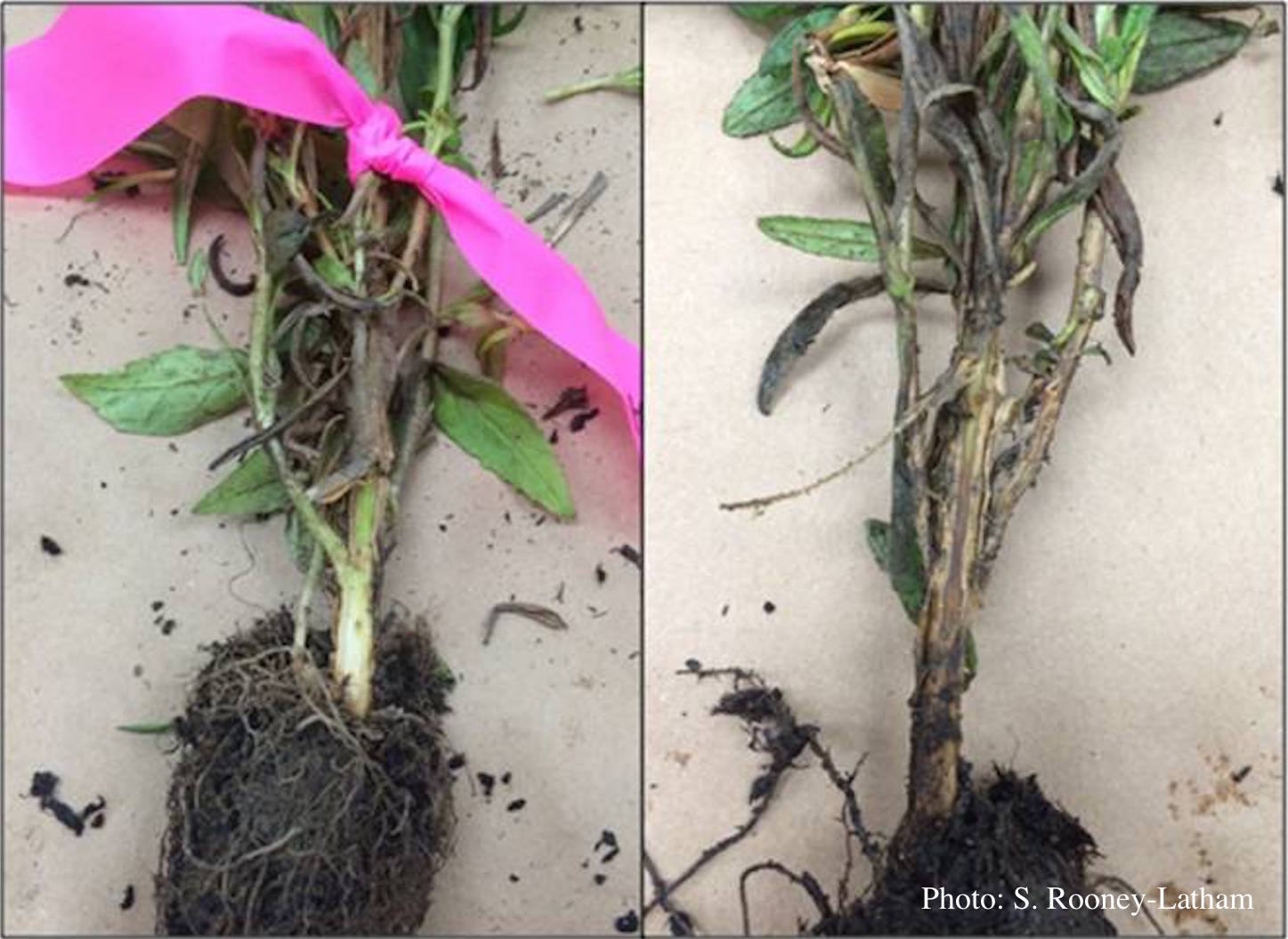 |
P. cactorum sporangia  P. cactorum sporangia |
P. alni basal canker on European Alder 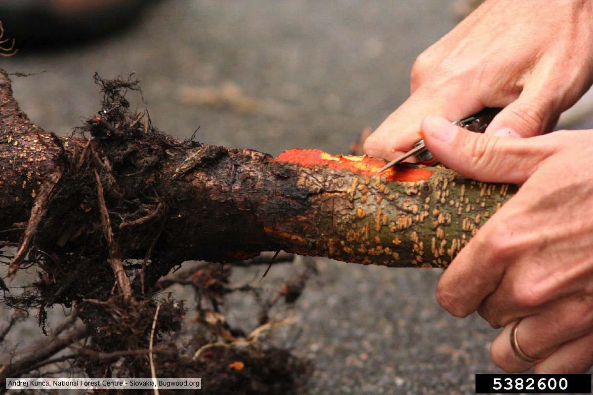 P. alni basal canker on European Alder (Alnus glutinosa) |
|
Chlamydospore of P. lateralis 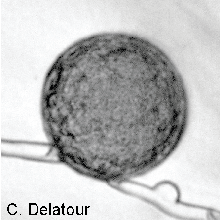 Laterally intercalary chlamydospore of Phytophthora lateralis |
P. arenaria sporangia  Globose papillate sporangia of Phytophthora arenaria on V8 agar flooded with soil extract. (Scale bar = 20 μm) |
P. cambivora on dead and dying chinquapin  Dead and dying chinquapin infected with P. cambivora |
|
P. cactorum bleeding canker 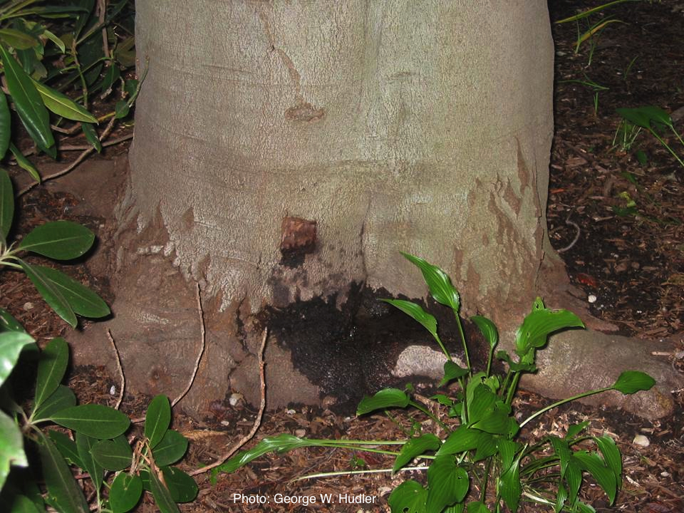 Bleeding canker on European beech (Fagus sylvatica) |
Dying Port Orford Cedar trees  |
P. austrocedrae colony morphology on Tomato juice agar with B sitosterol 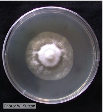 Colony morphology of P. austrocedrae at 16 ºC after 4 weeks on Tomato juice agar with B sitosterol |
|
P. cambivora colony morphology on PDA 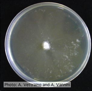 Appressed colony morphology at 14 days at 20°C on PDA |
P. palmivora symptoms on fruit  Brown rot on a lemon fruit caused by Phytophthora palmivora. |
P. lateralis on Port Orford cedar 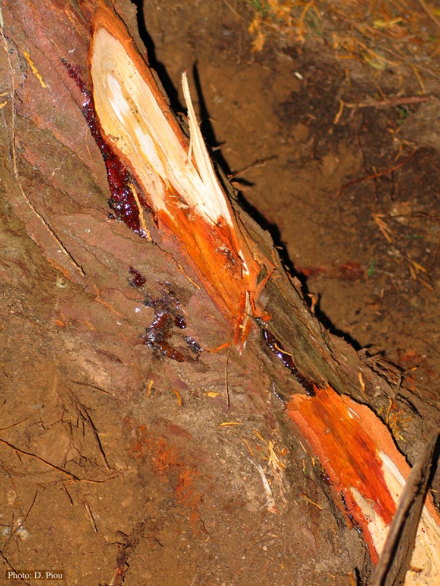 Small root lesions on Chaemacyparis lawsoniana |