

Phytophthora chlamydospora chlamydospore in agar. Bar is 20µm.
Photo Gallery
Site will be retired 9/1/2026
This site is no longer being developed and will be retired on September 1, 2026. Please contact us if you have any questions or would like to provide support to continue the project.
|
P. chlamydospora chlamydospore  |
P. alni in alder forest, Germany, with T. Jung 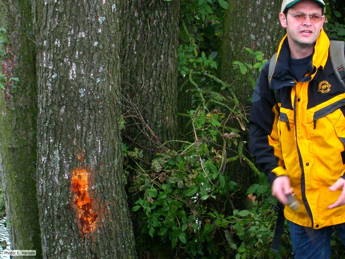 P. alni in alder forest, Germany, with T. Jung |
P. cryptogea colony morpholgy on PDA 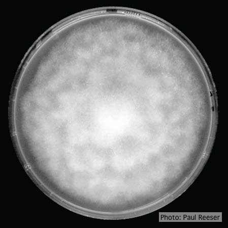 Colony morphology on PDA at 14 days |
|
P. palmivora sporangia 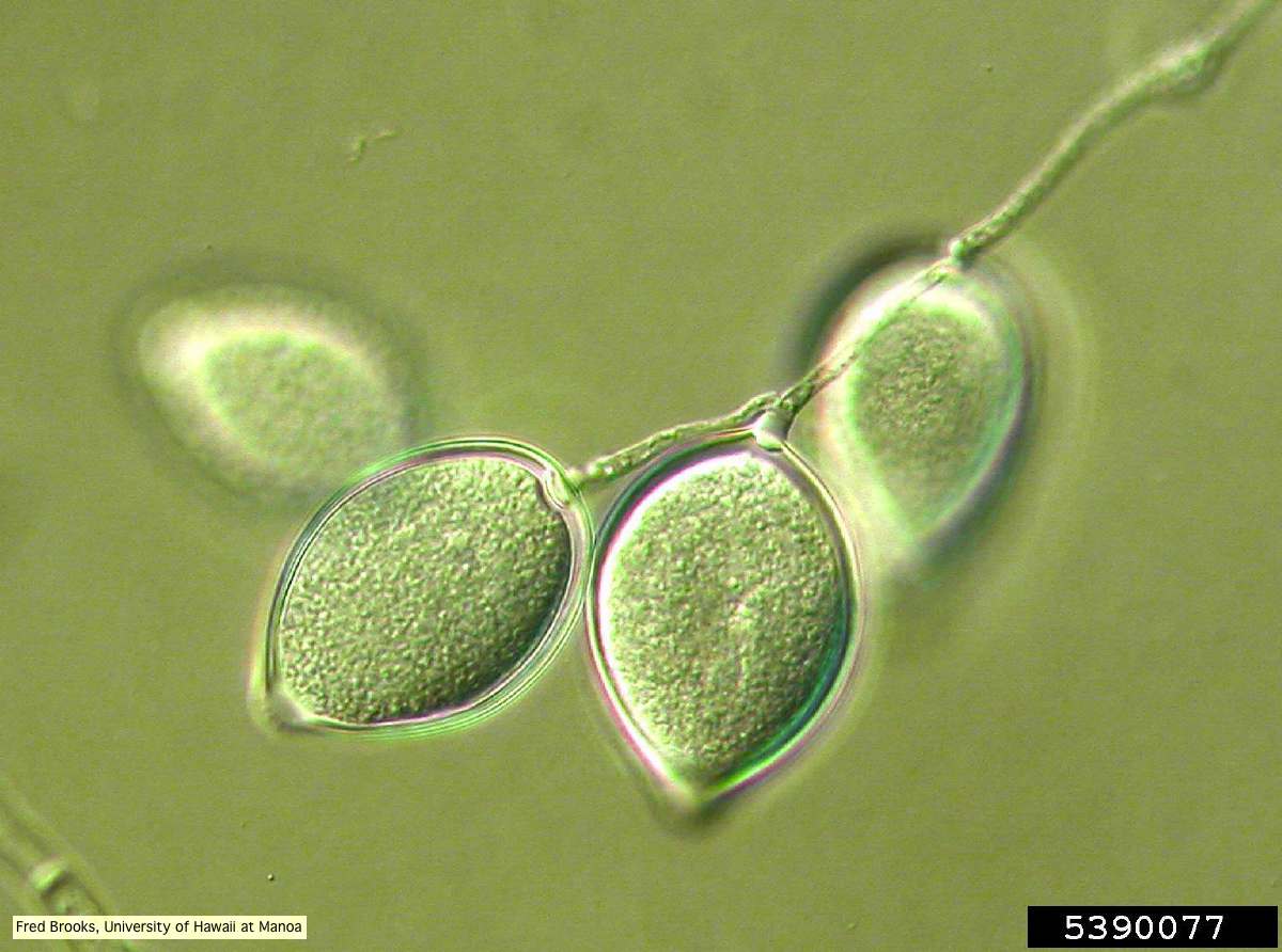 Sporangia (sporangiospores) showing sympodial branching |
P. tentaculata disease symptoms on California mugwort 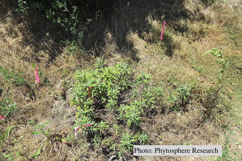 Outplanted California mugwort (Artemisia douglasiana) infected with P. tentaculata, 4.5 years after planting. Plant shows stunting and chlorosis. (P. cryptogea and P. lacustris were also baited from roots/soil of this plant). |
P. pinifolia sporangium 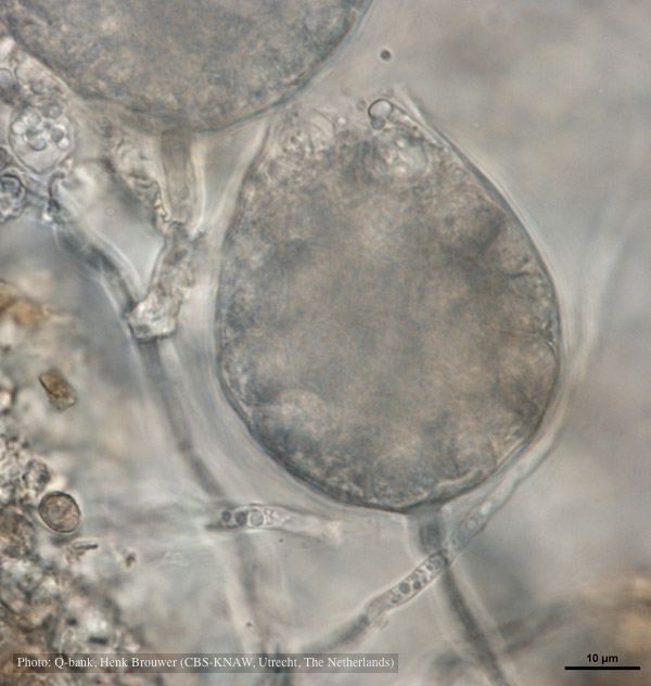 Cysts remain in sporangium after discharge, photo from Q-bank, used with permission |
|
P. agathidicida lesion on kauri tree 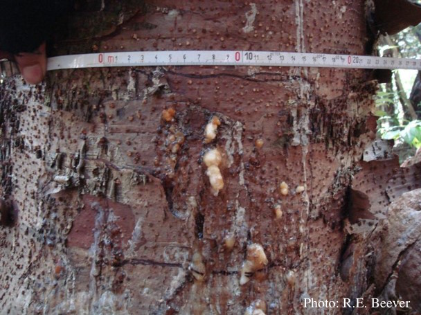 Close up of gum oozing out of lower trunk lesions of a young kauri tree at Maungaroa Ridge, Piha region of Waitakere Regional Park |
P. pinifolia on Pinus radiata 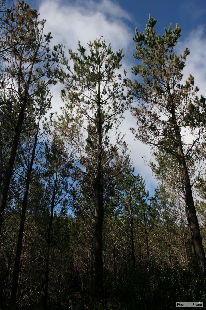 Pinus radiata impacted by DFP, note healthy new growth |
P. tentaculata disease symptoms on sticky monkey flower 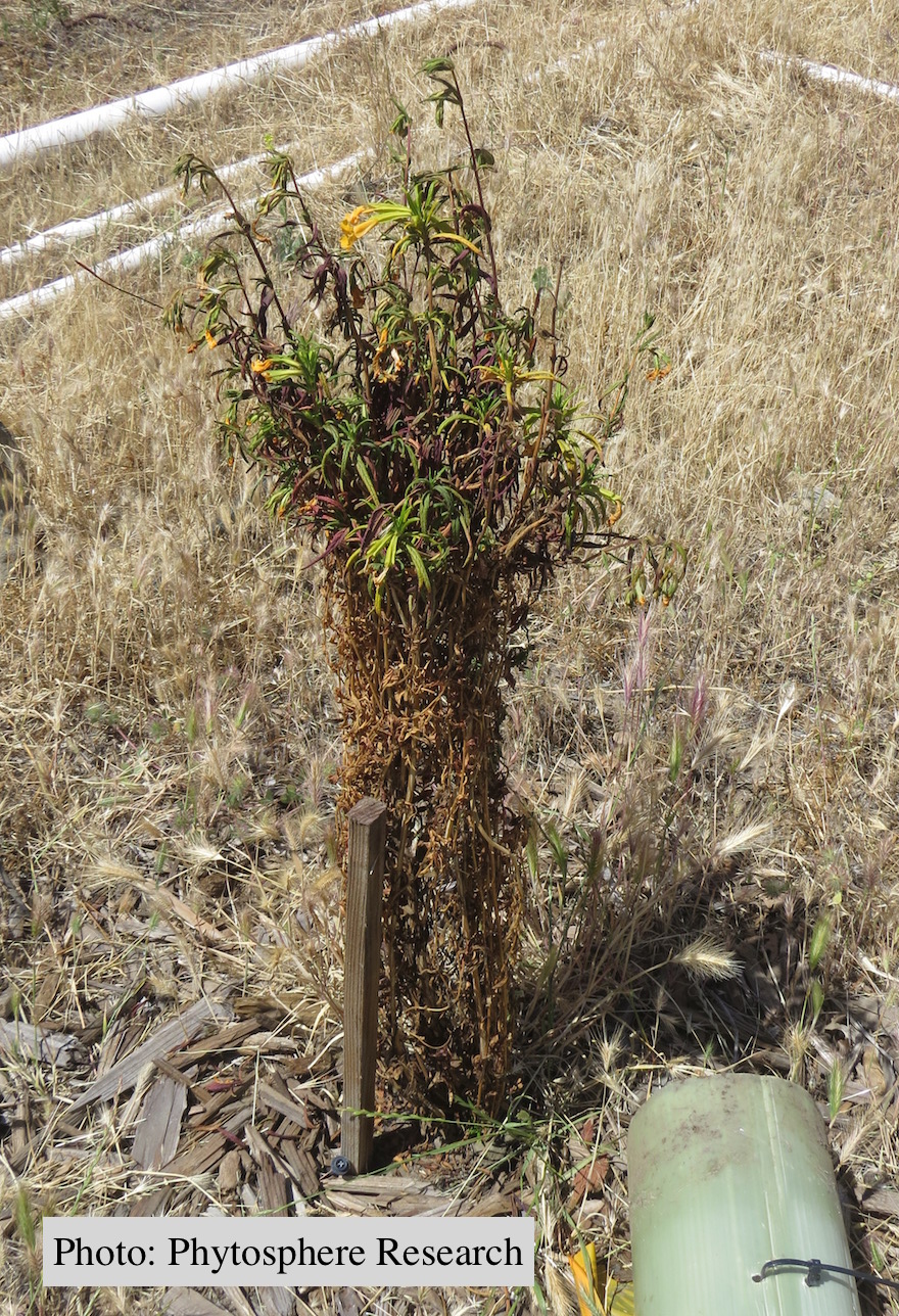 Outplanted sticky monkey flower (Diplacus aurantiacus) infected with P. |
|
P. pinifolia sporangia 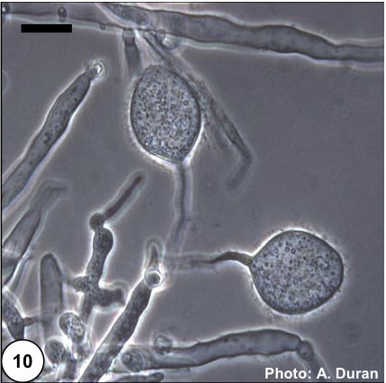 Non- papillate and caducous sporangia of Phytophthora pinifolia isolated from the infected P. radiata needles. |
P. kernoviae sporangium 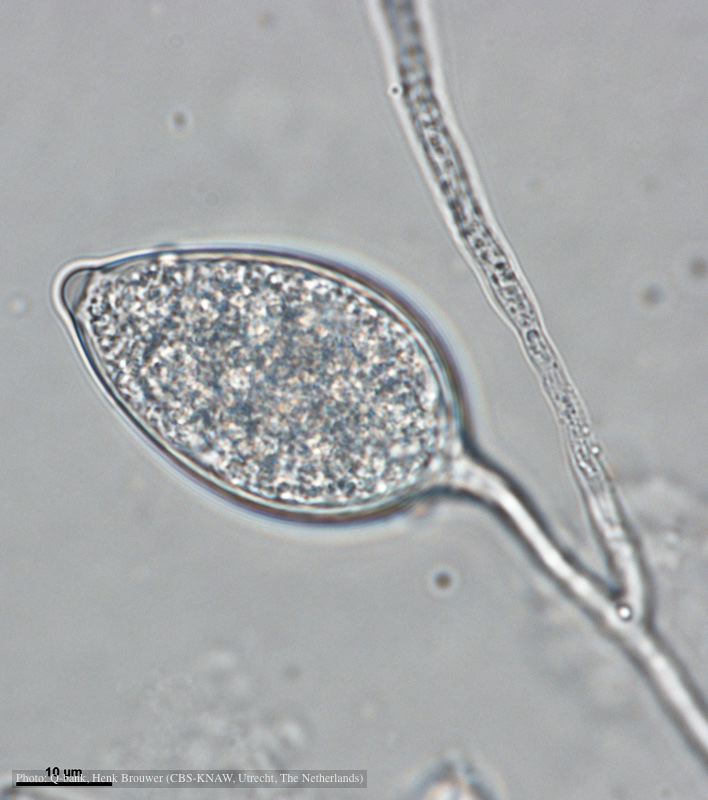 Papillate and caducous sporangium, photo from Q-bank, used with permission |
P. cactorum sporangium  P. cactorum sporangium |