

Ink disease impact in sweet chestnut forest in Italy
Photo Gallery
Site will be retired 9/1/2026
This site is no longer being developed and will be retired on September 1, 2026. Please contact us if you have any questions or would like to provide support to continue the project.
|
P. cambivora disease symptoms 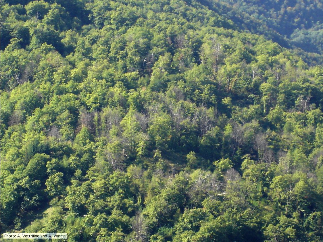 |
P. tentaculata sporangium 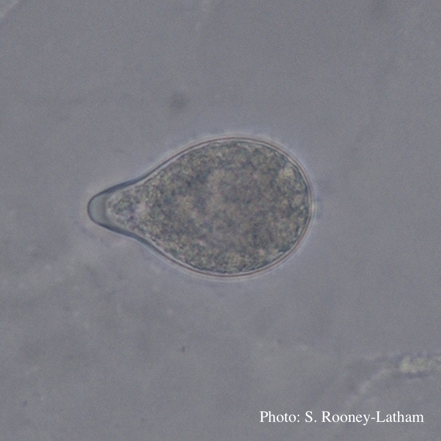 Papillate sporangium of P. tentaculata with an elongated neck or beak. |
P. cambivora sporangium with sympodial proliferation 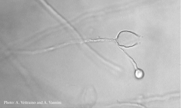 Empty sporagium showing sympodial proliferation |
|
P. austrocedrae semipapillate sporangium  P. austrocedrae - semipapillate sporangium with off-center attachment. |
P. siskiyouensis disease symptoms on Italian alder 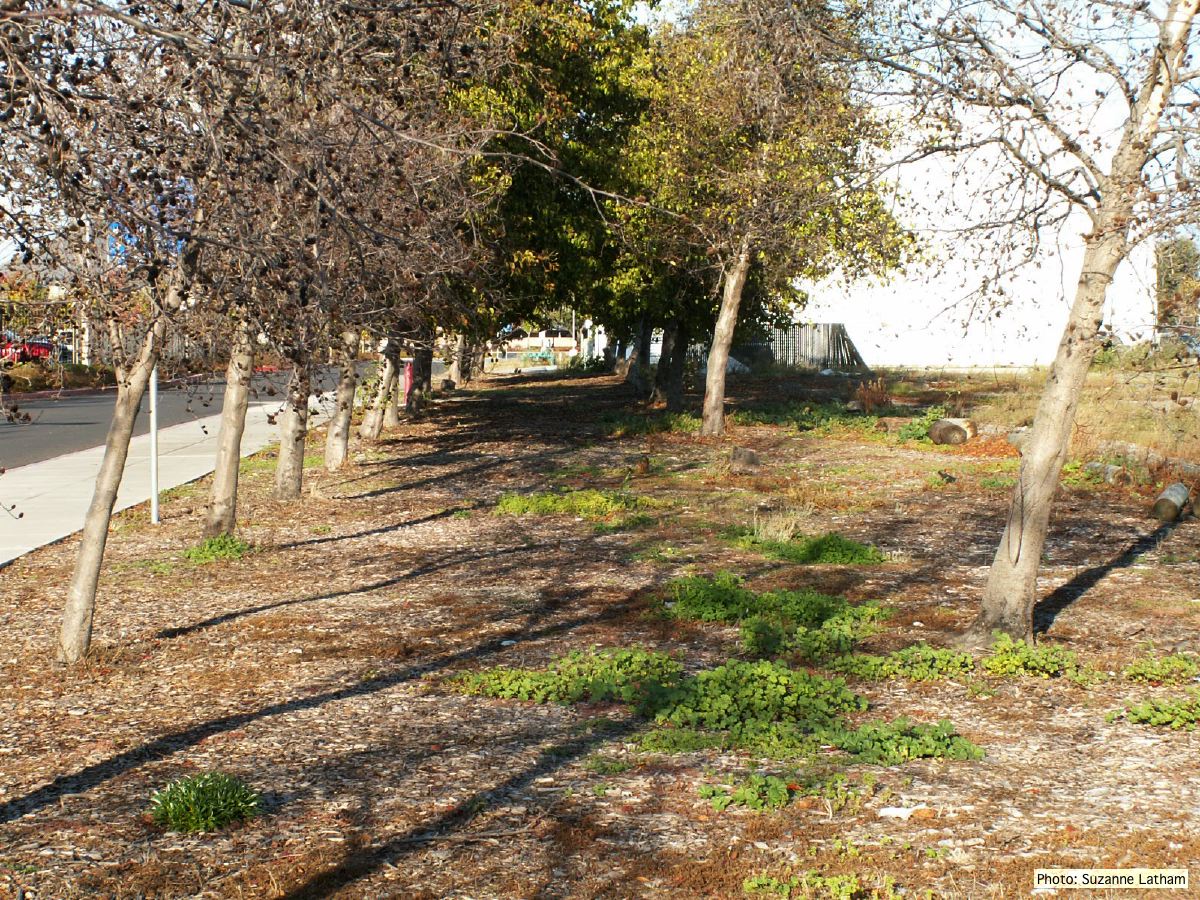 Grove of dying trees in a commercial landscape in Foster City, CA |
P. nicotianae chlamydospore 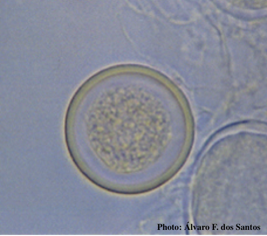 Globose chlamydospore (Fitopatol. bras. 2005) |
|
P. lateralis colony morphology on PDA 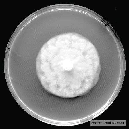 Growth morphology on PDA of Phytophthora lateralis |
P. kernoviae disease on European beech 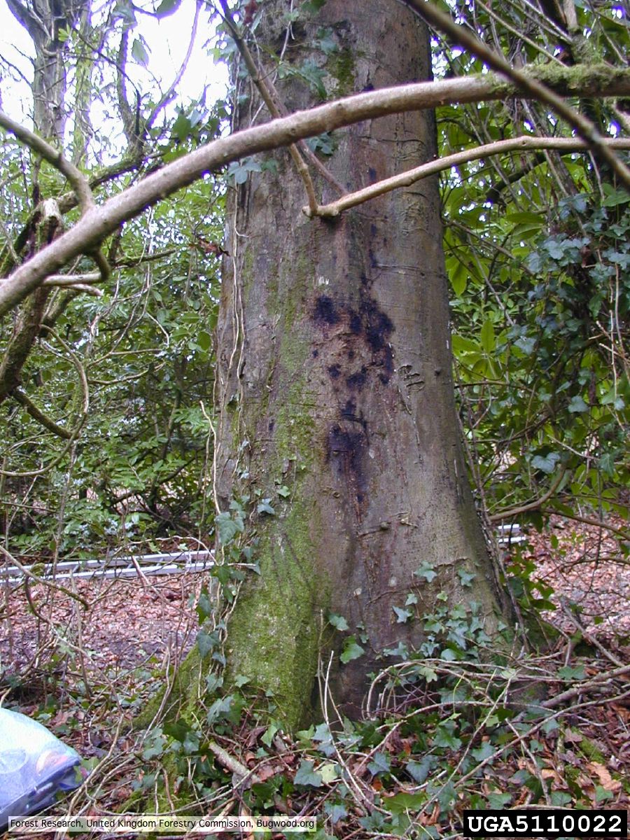 Bleeding lesion on trunk of Fagus sylvatica |
P. austrocedrae - hyphal swellings  Morphology of hyphae of Phytophthora austrocedrae, from Greslebin et al. 2007 |
|
P. boehmeriae chlamydospore 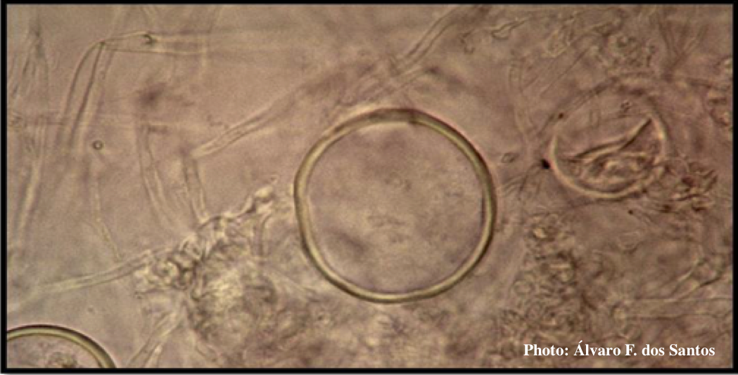 Globose chlamydospore of P. boehmeriae |
P. lateralis sporangia 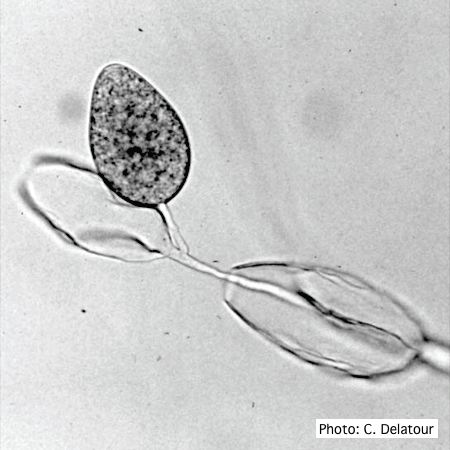 Sporangia of P. lateralis showing internal and external proliferation |
P. cinnamomi on Banksia 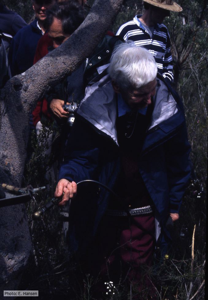 Gretna Weste injecting Banksia with phosphonate |