

Oogonium with paragynous antheridia applied close to the ogonial stalk.
Photo Gallery
Site will be retired 9/1/2026
This site is no longer being developed and will be retired on September 1, 2026. Please contact us if you have any questions or would like to provide support to continue the project.
|
P. megasperma oogonium 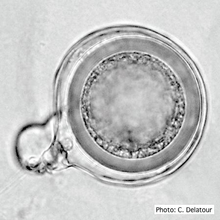 |
P. megakarya sporangium 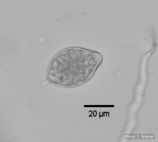 Caducous papillate sporangium of P. megakarya |
P. cambivora sporangium with sympodial proliferation 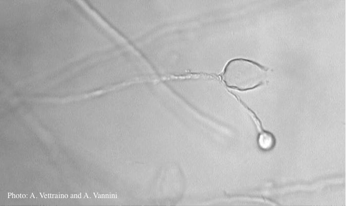 Empty sporagium showing sympodial proliferation |
|
P. cryptogea hyphal swellings 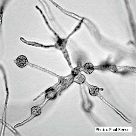 Cluster of small, angular to globose hyphal swellings formed in water |
P. kernoviae leaf wilt 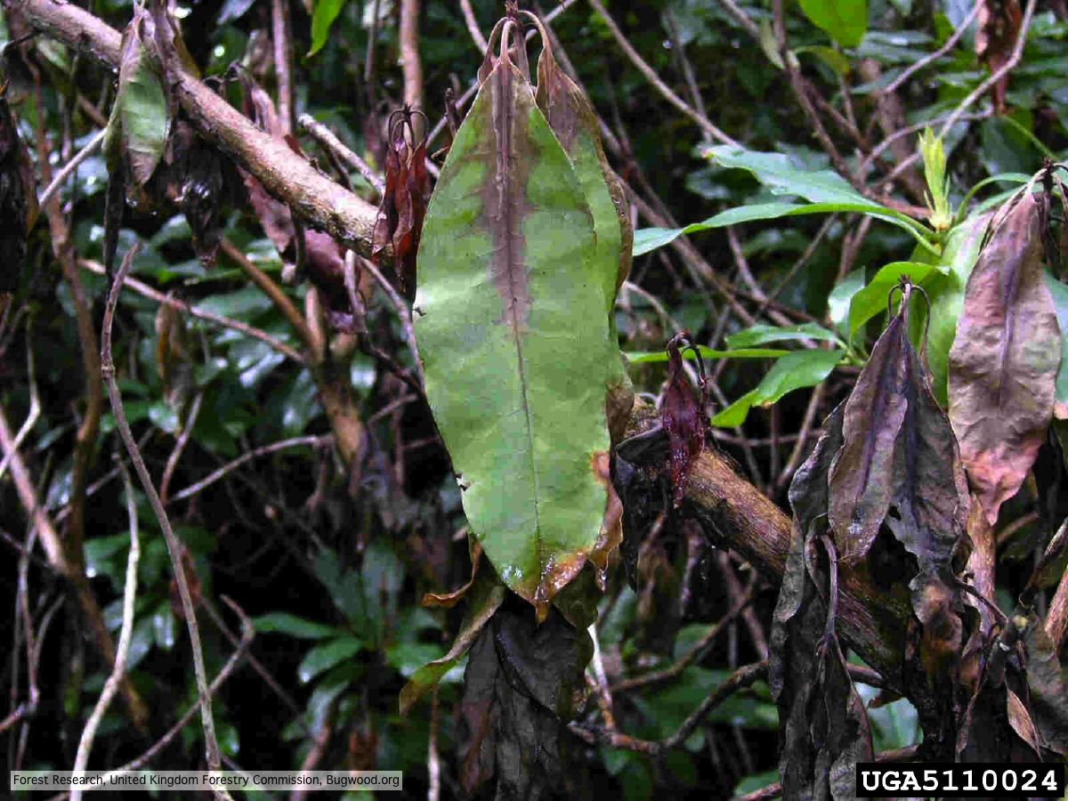 Wilted leaf of infected rhododendron |
P. austrocedrae necrotic lesion in phloem 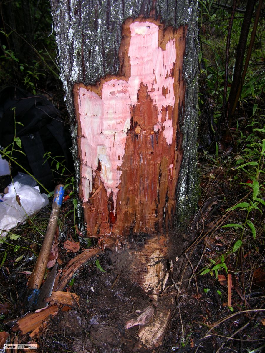 P. austrocedrae - necrotic lesion in phloem |
|
P. nicotianae chlamydospore 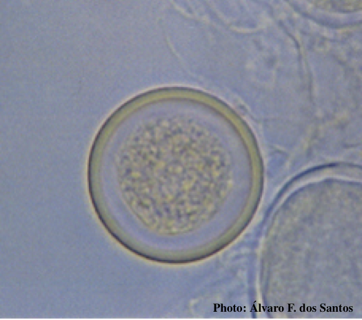 Globose chlamydospore (Fitopatol. bras. 2005) |
Growth of P. arenaria on CA  Colony morphology of Phytophthora arenaria after 7 days at 20°C on carrot agar |
P. pseudotsugae sporangia  Broadly ovoid, papillate sporangia in water |
|
P. kernoviae sporangia  Mycol.Res 109, 853-859; Figs 18-22. Regular, ovoid limoniform sporangia. Figs 23-26. Asymmetrical or sporangia |
P. cambivora sporangium 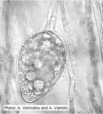 Ovoid non-papillate sporangia with well-rounded base |
P. pseudotsugae sporangium 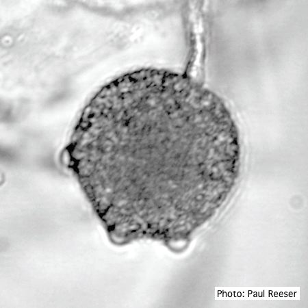 Broadly ovoid, papillate sporangium in water |