

Colony morphology of ex-holotype ICMP 17027 after 10-days incubation at 20°C in the dark
Photo Gallery
Site will be retired 9/1/2026
This site is no longer being developed and will be retired on September 1, 2026. Please contact us if you have any questions or would like to provide support to continue the project.
|
P. agathidicia growth on V8  |
P. pluvialis sporangia.  P. pluvialis sporangia on tape peel from infected Douglas-fir needle. |
P. tentaculata sporangium 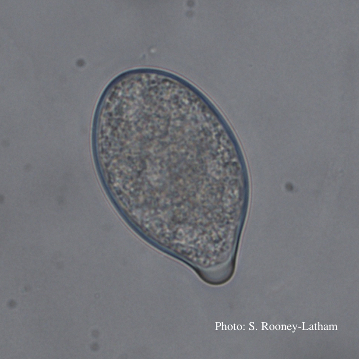 Papillate sporangium of P. tentaculata |
|
P. alni oogonia subspecies and variants 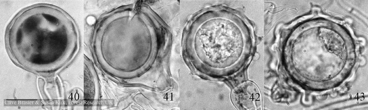 Fig. 40. P. alni subsp. uniformis. Fig. 41. P. alni subsp. multiformis German variant. Fig. 42. P. alni subsp. alni. Fig. 43. |
P. pluvialis oogonium and antheridium 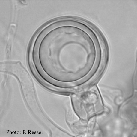 Oogonium and oospore with amphigynous antheridium |
P. pinifolia hyphal swellings  Spherical hyphal swelling with radiating hyphae (from Duran et al. 2008). Scale bar = 20 μm. |
|
P. palmivora sporangia, chlamydospores, hyphae 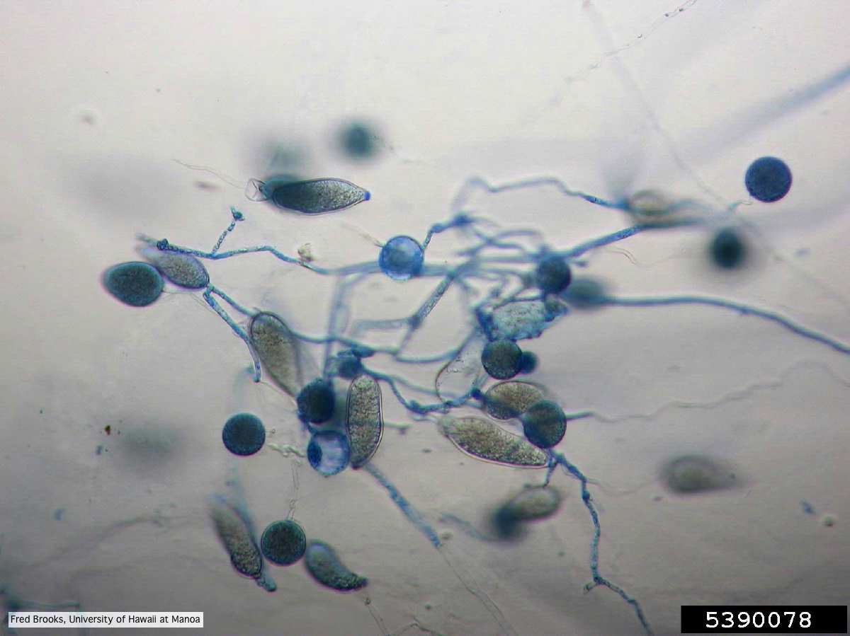 Sporangia (sporangiospores), chlamydospores, and hyphae stained with Cotton Blue |
P. pluvialis on Pinus radiata in New Zealand  Typical red needle cast symptoms along a twig. Lesions begin at the base of the needle which subsequently turns brown and is cast from the twig. |
P. ramorum sporangium 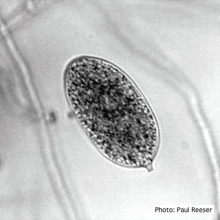 P. ramorum sporangium |
|
P. pinifolia coenocytic hyphae 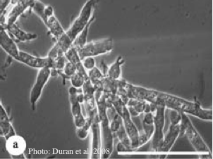 Coenocytic hyphae (from Duran et al. 2008). Scale bar = 20 μm. |
P. lateralis colony morphology on PDA 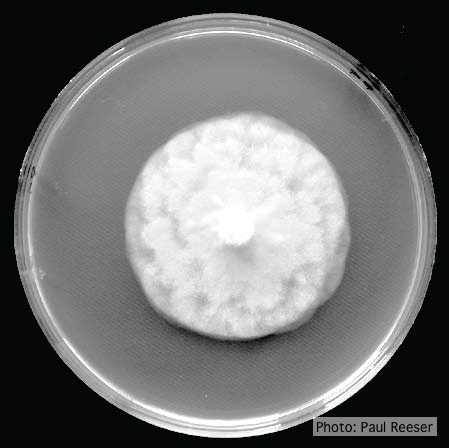 Growth morphology on PDA of Phytophthora lateralis |
P. agathadicida disease symptom 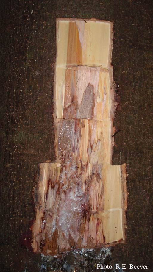 Excavated lesion, with outer bark removed showing extent of disease-front |