

Nursery grown California mugwort plant (Artemisia douglasiana) infected with P. tentaculata and exhibiting severe root and crown rot
Photo Gallery
Site will be retired 9/1/2026
This site is no longer being developed and will be retired on September 1, 2026. Please contact us if you have any questions or would like to provide support to continue the project.
|
P. tentaculata disease symptoms on California mugwort  |
P. cambivora sporangium 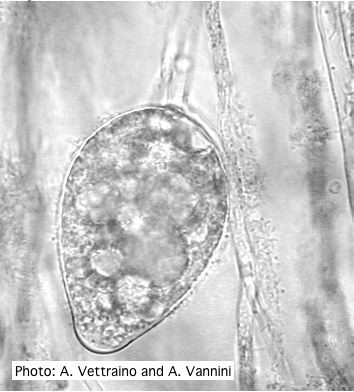 Ovoid non-papillate sporangia with well-rounded base |
Growth morphology on V8 of P. lateralis 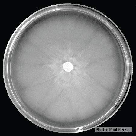 Colony morphology on V8 at 14 days |
|
P. austrocedrae necrotic lesion in phloem 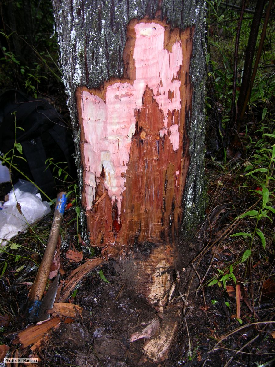 P. austrocedrae - necrotic lesion in phloem |
P. palmivora oogonium 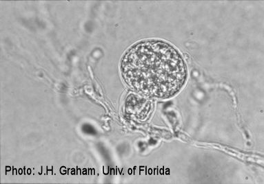 P. palmivora oogonium |
P. pinifolia on Pinus radiata 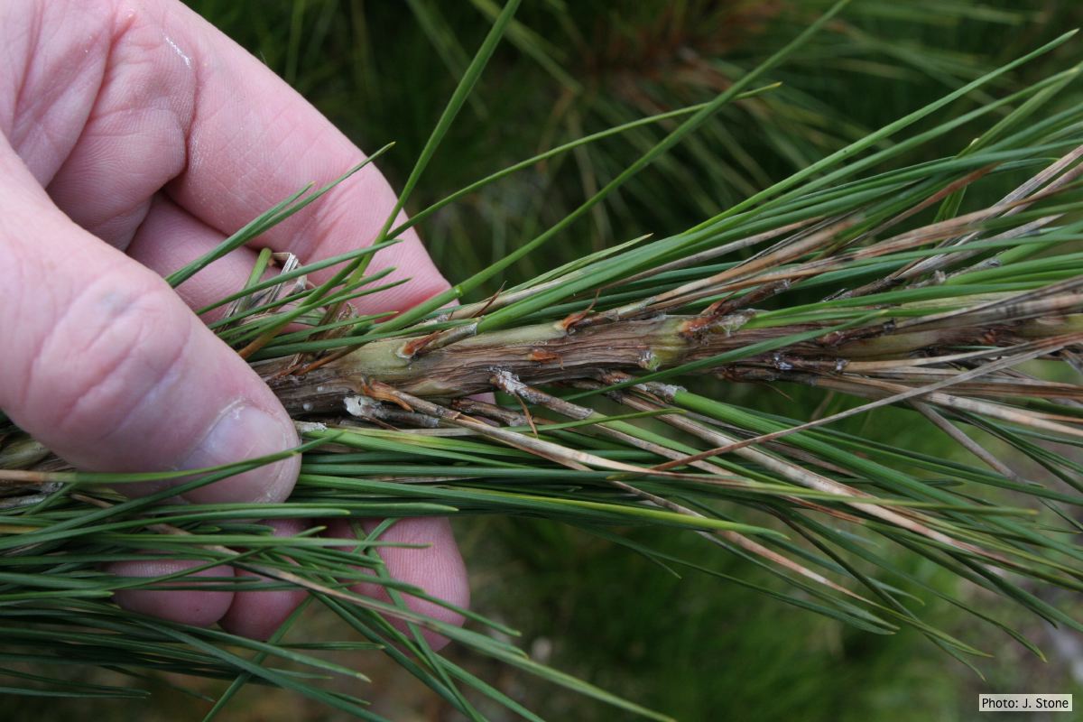 Pinus radiata needles, note “black line” symptom near needle bases |
|
P. boehmeriae oogonia  Oogonia and oospores with amphigynous antheridia |
P. pinifolia coenocytic hyphae 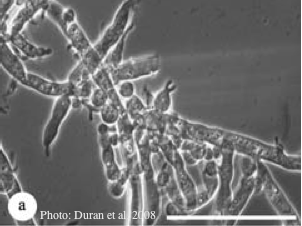 Coenocytic hyphae (from Duran et al. 2008). Scale bar = 20 μm. |
P. tentaculata disease symptoms on California mugwort 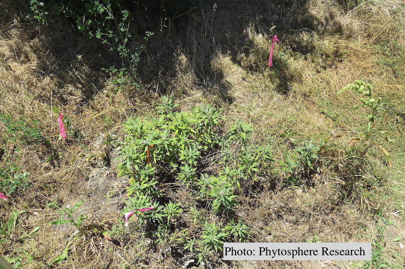 Outplanted California mugwort (Artemisia douglasiana) infected with P. tentaculata, 4.5 years after planting. Plant shows stunting and chlorosis. (P. cryptogea and P. lacustris were also baited from roots/soil of this plant). |
|
P. agathidicida lesion on kauri tree 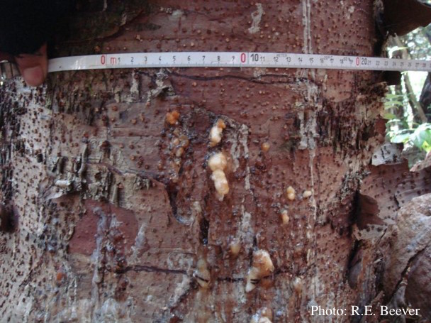 Close up of gum oozing out of lower trunk lesions of a young kauri tree at Maungaroa Ridge, Piha region of Waitakere Regional Park |
P. agathidicia sporangia 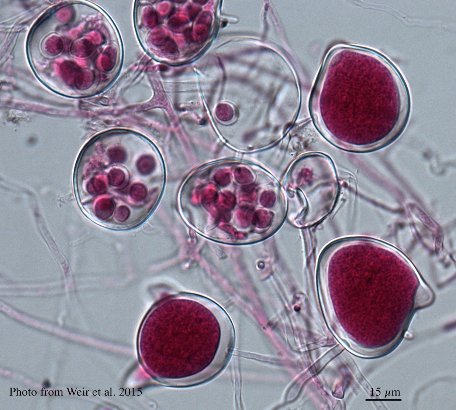 Differentiation of the cytoplasm within papillate sporangia into acid fuchsin stained zoospores |
P. agathidicida oospores in planta  Oospores in the roots of kauri seedlings inoculated with P. agathidicida. The root has been cleared with potassium hydroxide and bleached with peroxide before being stained with trypan blue (scale bar =100 µm). |