

Bleeding canker, European beech, OSU campus
Photo Gallery
Site will be retired 9/1/2026
This site is no longer being developed and will be retired on September 1, 2026. Please contact us if you have any questions or would like to provide support to continue the project.
|
P. cambivora symptoms  |
P. pinifolia hyphal growth 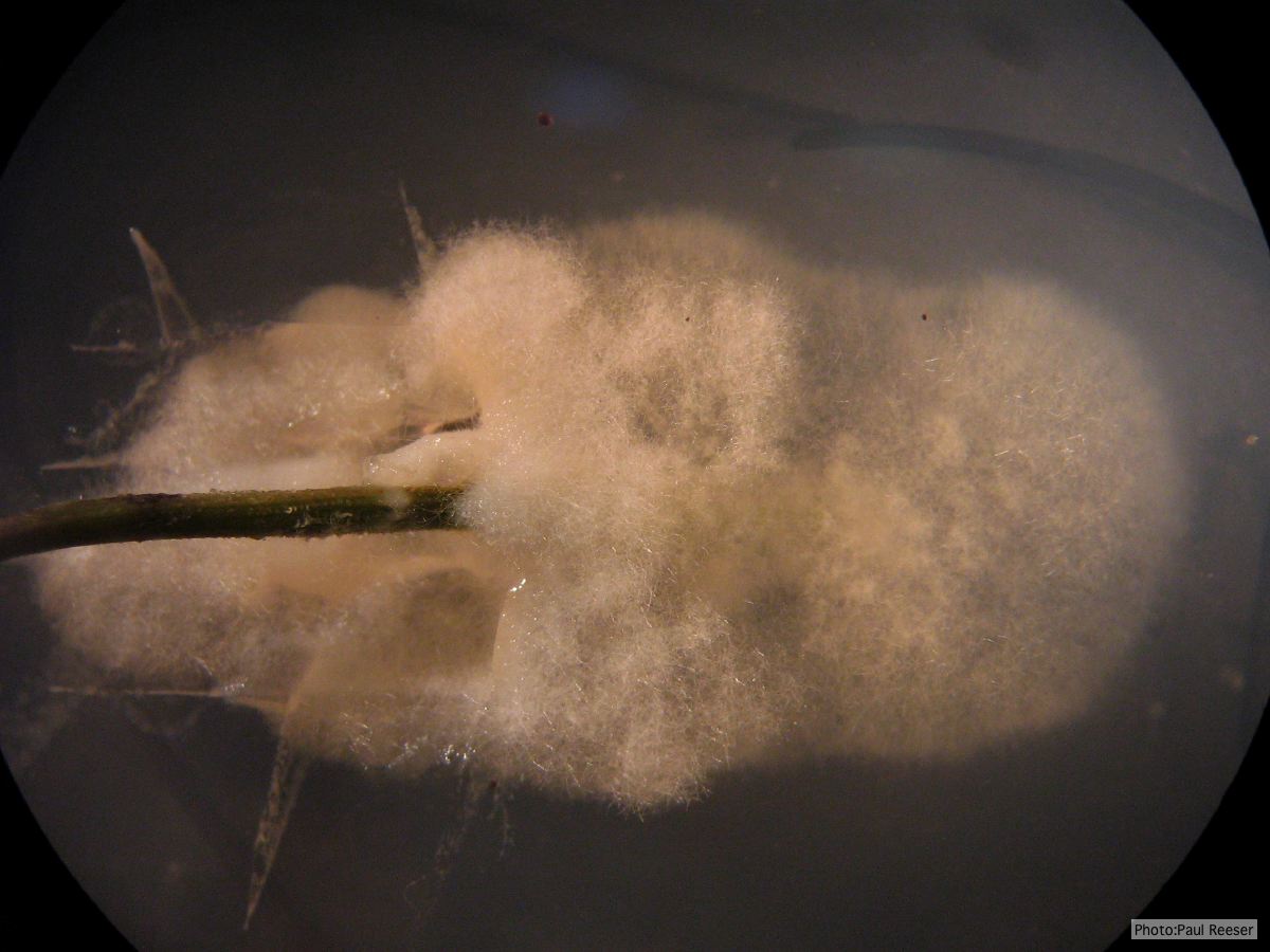 P. pinifolia pathogen growing from infected needle on selective agar |
P. kernoviae disease on beech 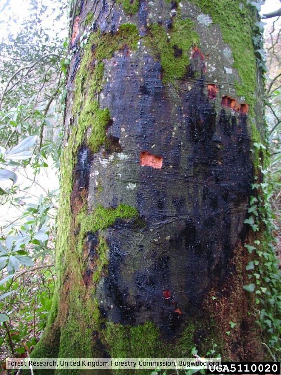 External lesion; 14 November 2003 |
|
P. frigida oogonium  Oogonium and oospore with amphigynous antheridium |
P. cambivora colony morphology on PDA  Cottony colony morphology at 14 days at 20°C on PDA |
P. cambivora oogonium 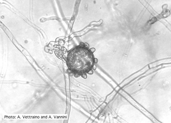 Bullate oogonium with amphyginous antheridium |
|
Phytophthora cactorum disease symptoms on English walnut 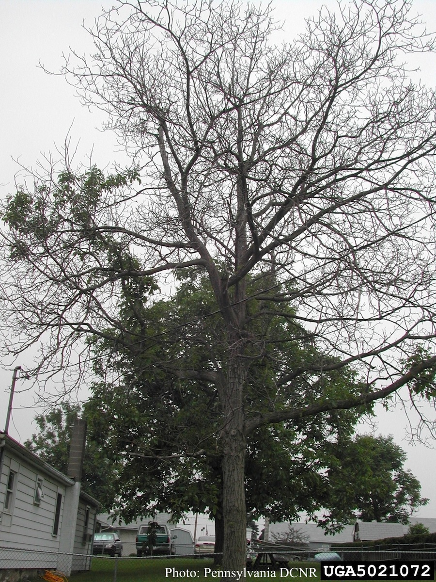 Mortality of Juglans regia caused by Phytophthora cactorum. |
P. pseudotsugae amphigynous oogonium 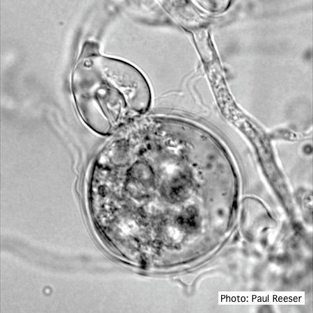 P. pseudotsugae oogonium with amphigynous antheridia |
P. cambivora oogonium 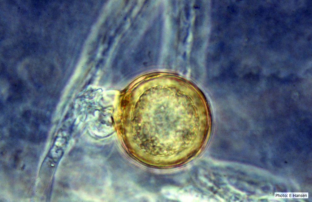 P. cambivora oogonium with antheridium |
|
P. lateralis on Port Orford cedar  Root lesions on Chaemacyparis lawsoniana |
P. megakarya disease symptoms on Theobroma cacao fruit 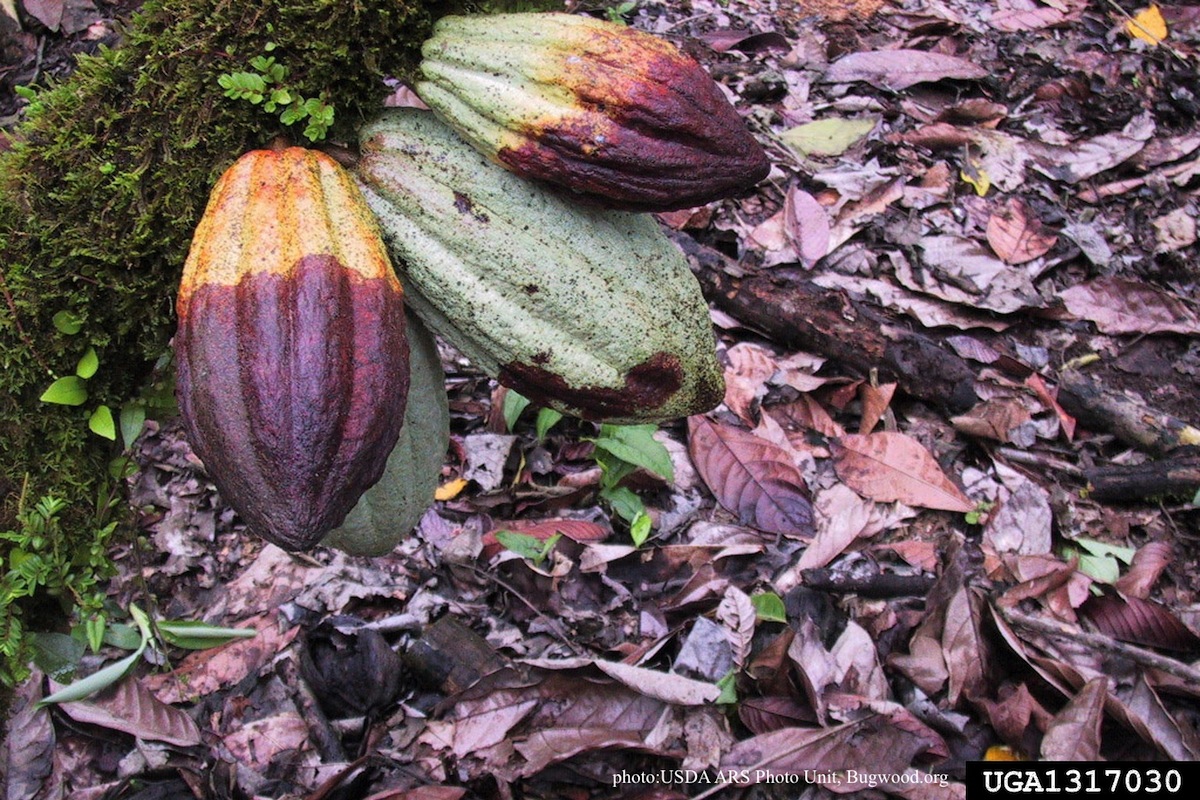 Disease symptoms on a cocoa pod |
P. chlamydospora chlamydospore 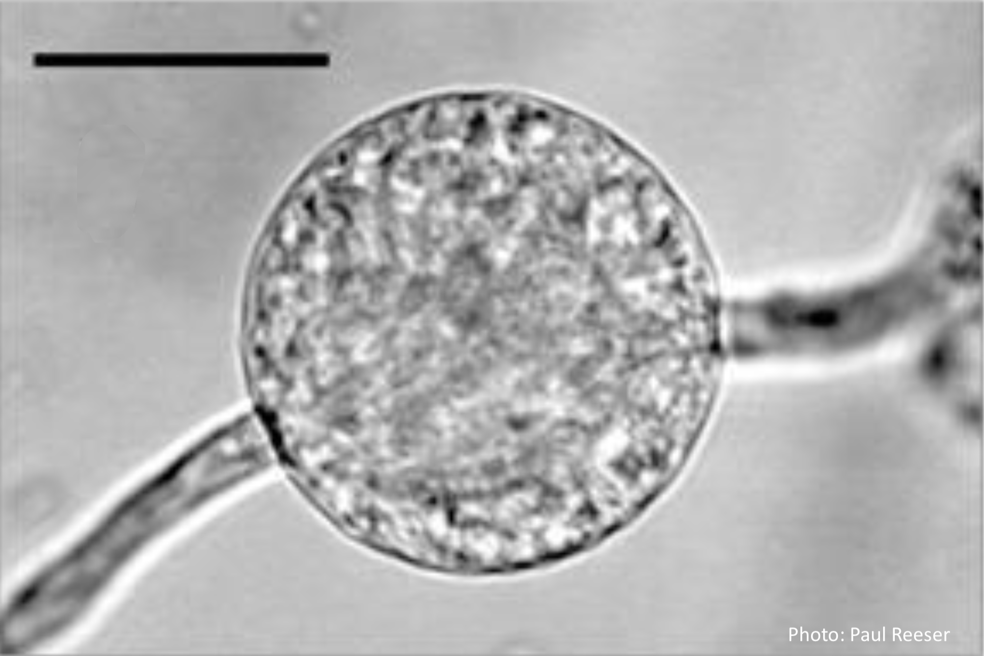 Phytophthora chlamydospora chlamydospore in agar. Bar is 20µm. |