

Dead Chamaecyparis lawsoniana, BLM Roseburg District in Oregon
Photo Gallery
Site will be retired 9/1/2026
This site is no longer being developed and will be retired on September 1, 2026. Please contact us if you have any questions or would like to provide support to continue the project.
|
Dead Port Orford Cedar 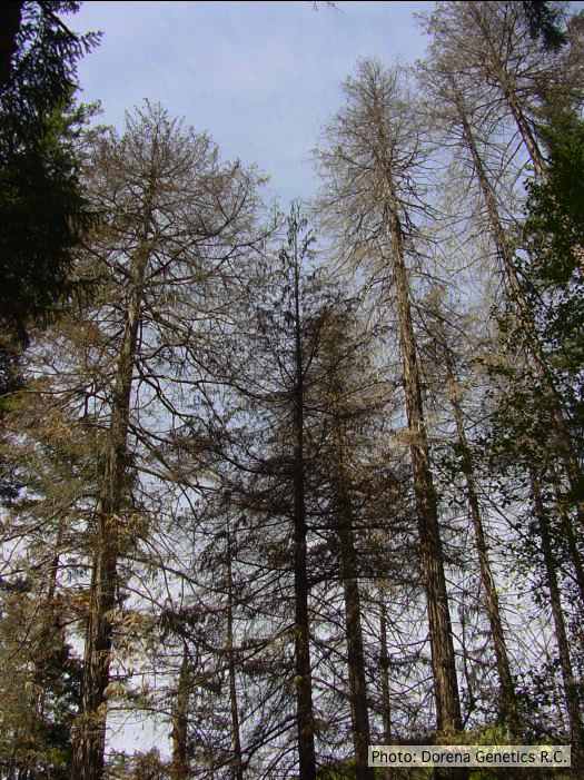 |
P. cactorum bleeding canker 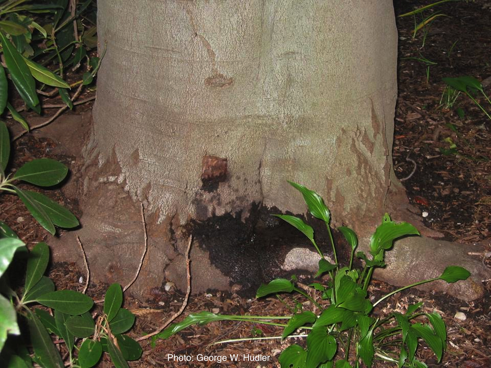 Bleeding canker on European beech (Fagus sylvatica) |
P. nicotianae colony morphology on V8 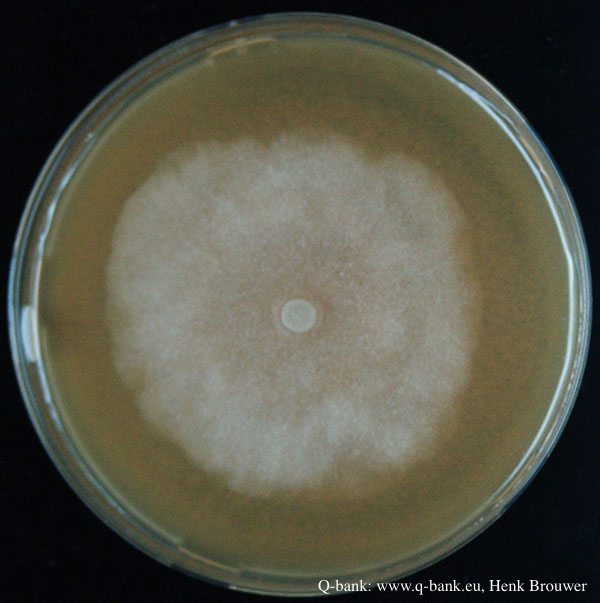 Phytophthora nicotianae CBS 321.49 V8 after 7 days at 24 degrees. Photo from Q-bank: www.q-bank.eu, Henk Brouwer (CBS-KNAW, Utrecht, The Netherlands) |
|
P. cryptogea colony morpholgy on PDA 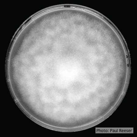 Colony morphology on PDA at 14 days |
P. kernoviae leaf wilt 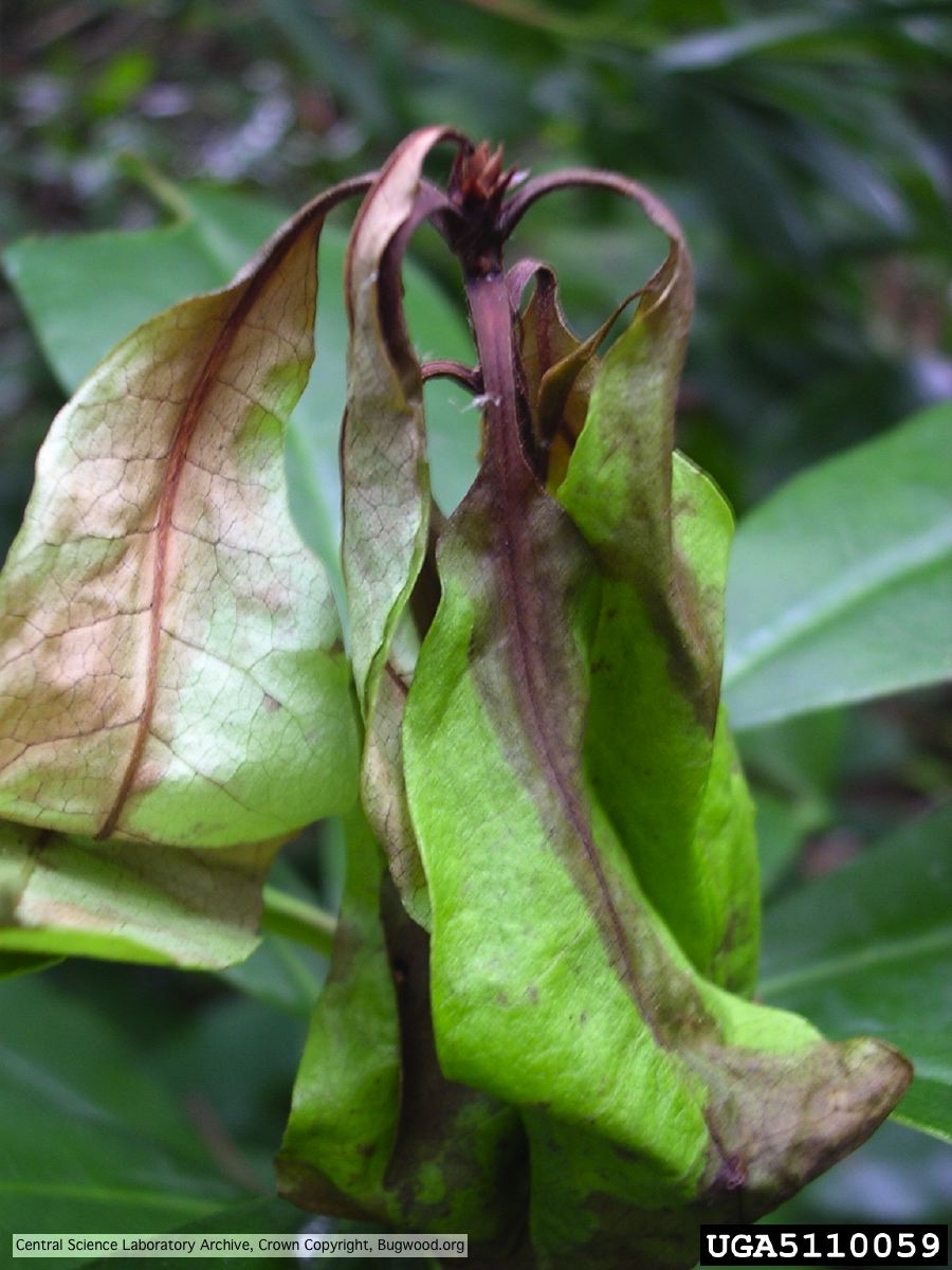 Necrosis of rhododendron leaves. |
P. nicotianae sporangia  P. nicotianae overview of sporangia 40x. Photo from Q-bank: www.q-bank.eu, Henk Brouwer (CBS-KNAW, Utrecht, The Netherlands) |
|
P. megakarya chlamydospore 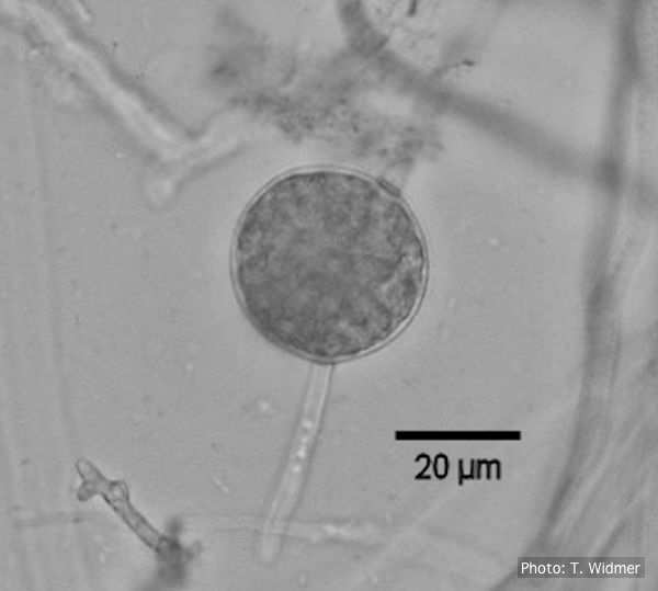
Terminal chlamydospore of P. megakarya
|
P. boehmeriae oogonium 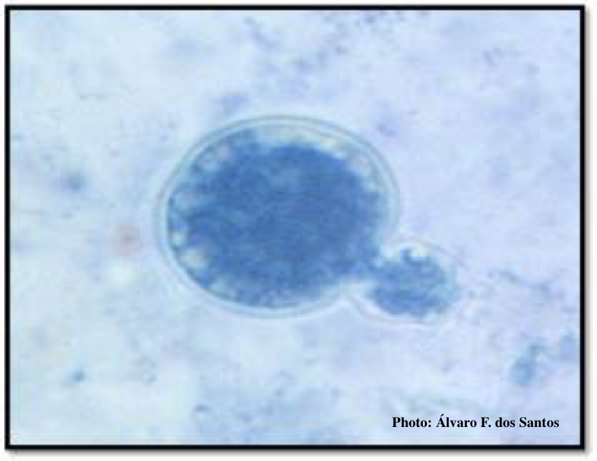 Oogonia and oospores with amphigynous antheridia |
P. kernoviae disease on beech 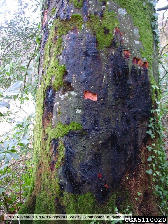 External lesion; 14 November 2003 |
|
P. siskiyouensis disease symptoms on Italian alder 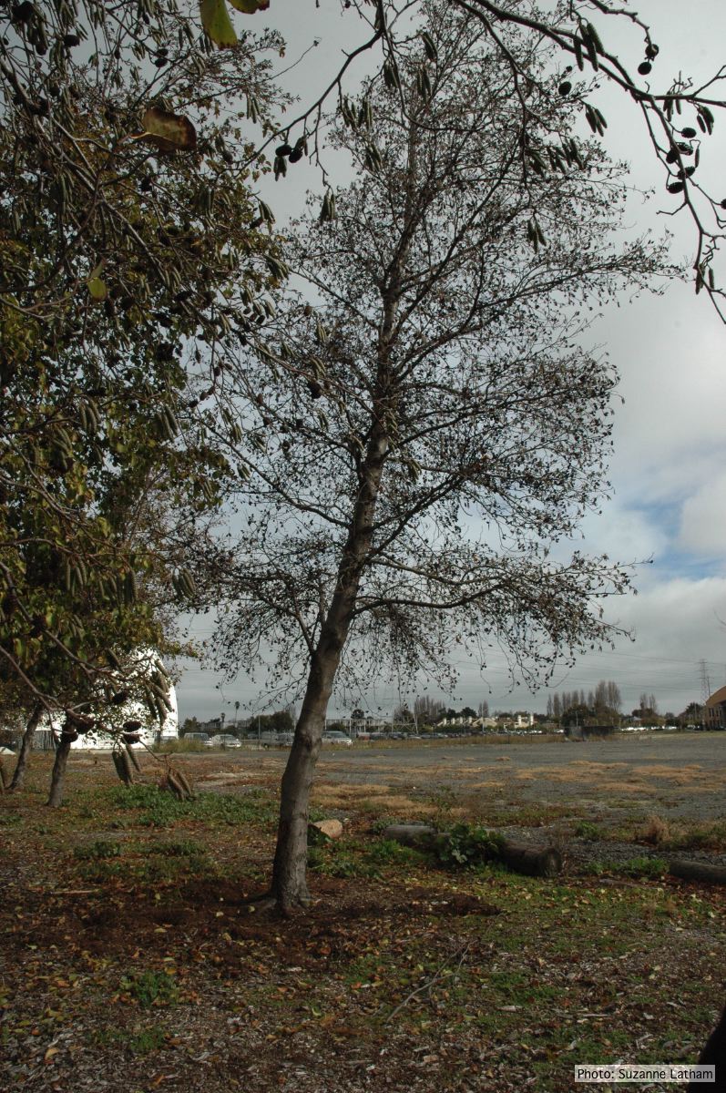 Phytophthora collar rot on Italian alder trees: standing, dead tree |
P. siskiyouensis oogonium with paragynous antheridium  P. siskiyouensis oogonium with paragynous antheridium |
P. austrocedrae - Mal del ciprés, stages of decline 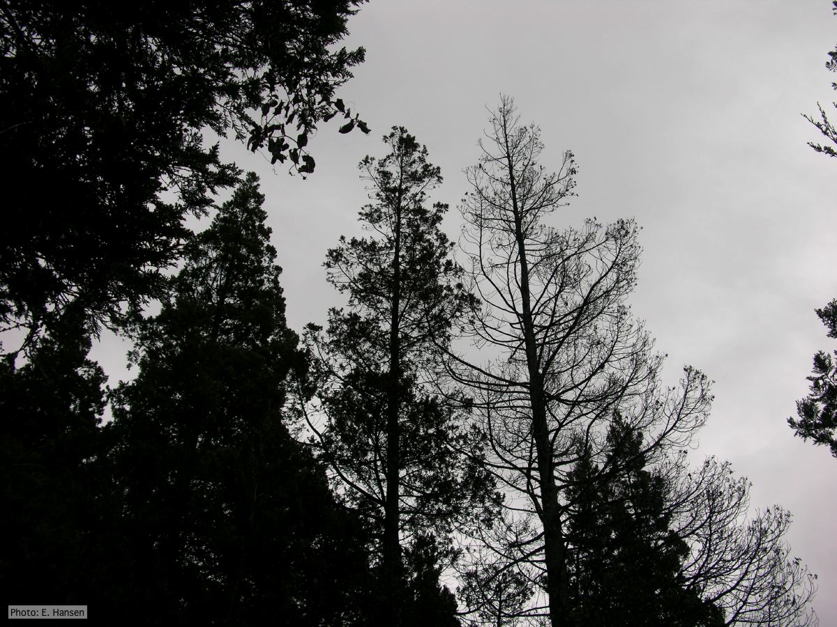 Mal del ciprés, stages of decline |