P. megakarya oogonia, oospore, and antheridium


|
P. megakarya oospore
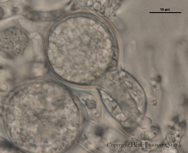
P. megakarya oogonia, oospore, and antheridium
|
P. pinifolia colony morphology on V8 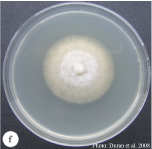 Colony morphology of P. pinifolia at 20°C on V8 after 3 weeks. From Duran et al. 2008 |
P. agathadicida disease symptom 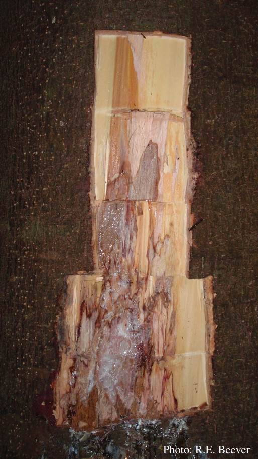 Excavated lesion, with outer bark removed showing extent of disease-front |
|
P. cambivora sporangium with internal extended proliferation  Empty sporagia showing internal nested and extended proliferation |
P. cryptogea hyphal swellings 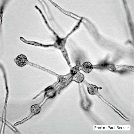 Cluster of small, angular to globose hyphal swellings formed in water |
P. pluvialis symptoms on Douglas-fir  Red needle cast symptoms on Douglas-fir in western Oregon, 2015 |
|
P. ramorum sporangium  Deciduous sporangium, photo from Q-bank, used with permission |
Dead Port Orford Cedar 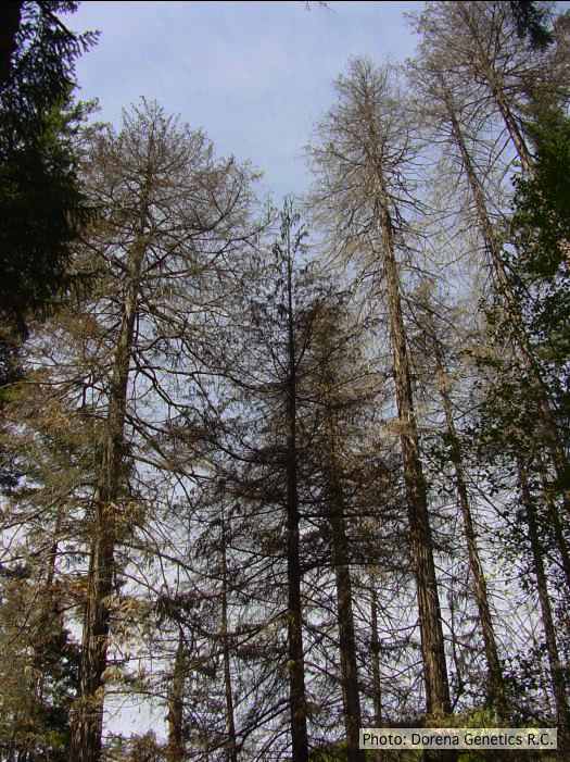 Dead Chamaecyparis lawsoniana, BLM Roseburg District in Oregon |
P. kernoviae sporangium 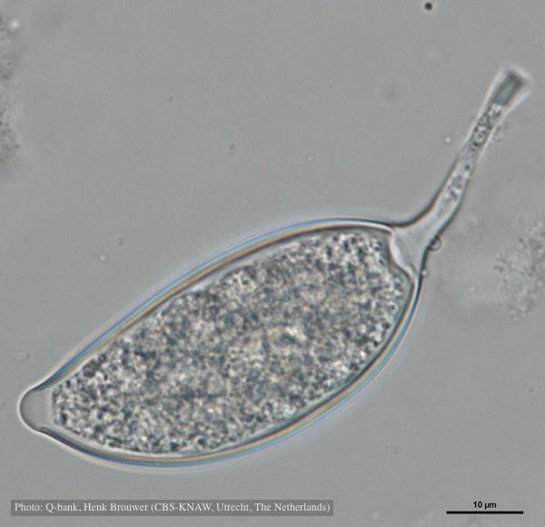 Asymmetrical sporangium, photo from Q-bank, used with permission |
|
P. cryptogea sporangium  Ovoid non-papillate sporangia in water. |
Dying Port Orford Cedar trees  |
Dead and healthy Port-Orford cedar seedlings  Port-Orford-cedar seedlings planted to test for Phytophthora lateralis resistance at the Dorena Genetic Resource Center |