

Nursery grown California mugwort plant (Artemisia douglasiana) infected with P. tentaculata and exhibiting severe root and crown rot
Photo Gallery
Site will be retired 9/1/2026
This site is no longer being developed and will be retired on September 1, 2026. Please contact us if you have any questions or would like to provide support to continue the project.
|
P. tentaculata disease symptoms on California mugwort  |
P. katsurae disease symptoms 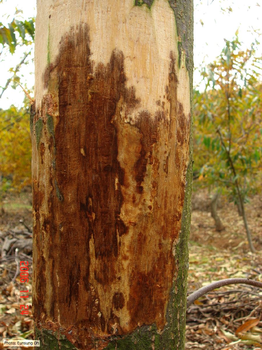 Infected chestnut (Castanea) with girdling canker |
P. pseudosyringae sporangia  Ovoid, semipapillate sporangia showing sympodial development of sporangiophore |
|
Phytophthora cactorum disease symptoms on English walnut  Dieback of Juglans regia caused by Phytophthora cactorum |
P. frigida symptoms 4 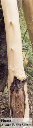 Black wattle timber with symptoms of gummosis |
Phytophthora cactorum disease symptoms on English walnut 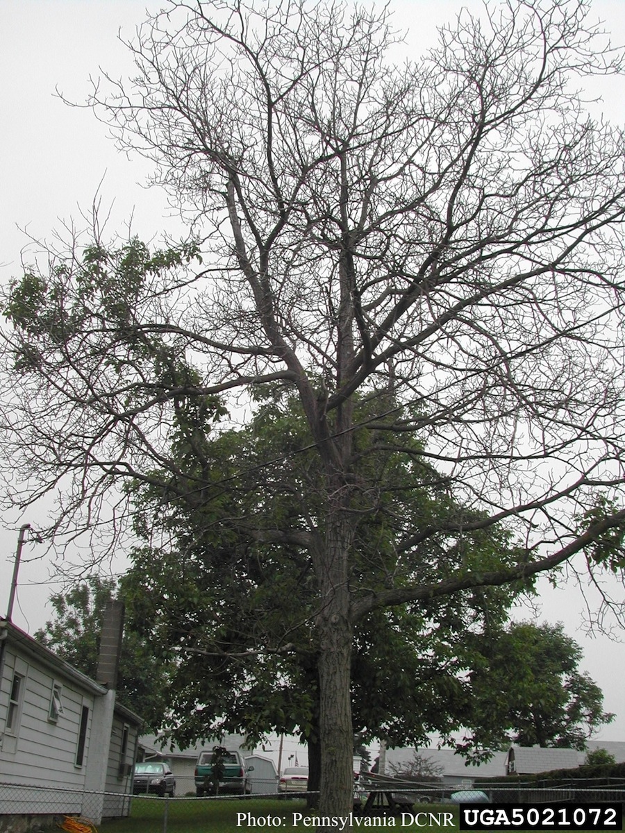 Mortality of Juglans regia caused by Phytophthora cactorum. |
|
P. pluvialis colony morphology on carrot agar  Colony morphology on carrot agar at 20 days |
P. palmivora symptoms on fruit  Brown rot on a lemon fruit caused by Phytophthora palmivora. |
Dead and healthy Port-Orford cedar seedlings  Port-Orford-cedar seedlings planted to test for Phytophthora lateralis resistance at the Dorena Genetic Resource Center |
|
P. pluvialis symptoms on Douglas-fir needles 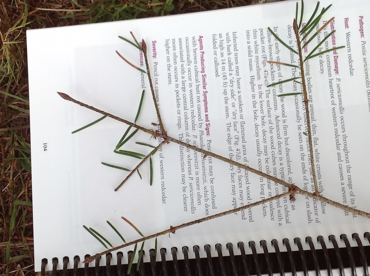 Symptoms of red needle cast on Douglas-fir needles |
P. nicotianae colony morphology on CMA  Phytophthora nicotianae CBS 321.49 CMA after 7 days at 24 degrees. Photo from Q-bank: www.q-bank.eu, Henk Brouwer (CBS-KNAW, Utrecht, The Netherlands) |
P. kernoviae colony morphology  From Mycol.Res 109, 853-859; growth on CA under different conditions |