

Colony morphology on PDA at 14 days
Photo Gallery
Site will be retired 9/1/2026
This site is no longer being developed and will be retired on September 1, 2026. Please contact us if you have any questions or would like to provide support to continue the project.
|
P. nemorosa colony morphology on PDA  |
P. lateralis sporangia  Sympodial sporangiophore with external proliferation |
P. alni symptoms on European Alder  Mature, riparian common alder (A. glutinosa) stand heavily impacted by root and collar rot caused by P. alni |
|
P. palmivora sporangia 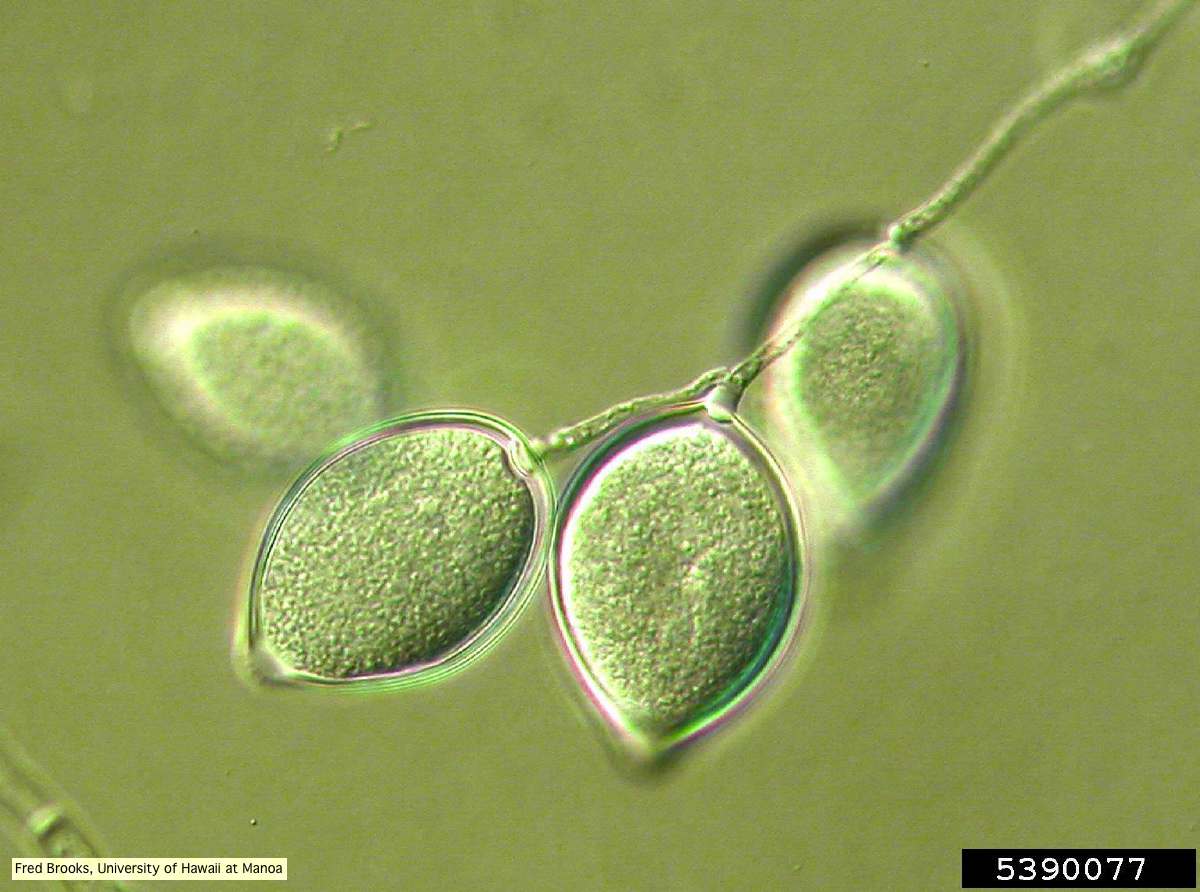 Sporangia (sporangiospores) showing sympodial branching |
P. megakarya chlamydospore 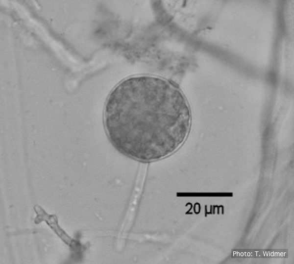
Terminal chlamydospore of P. megakarya
|
P. pluvialis symptoms on Douglas-fir 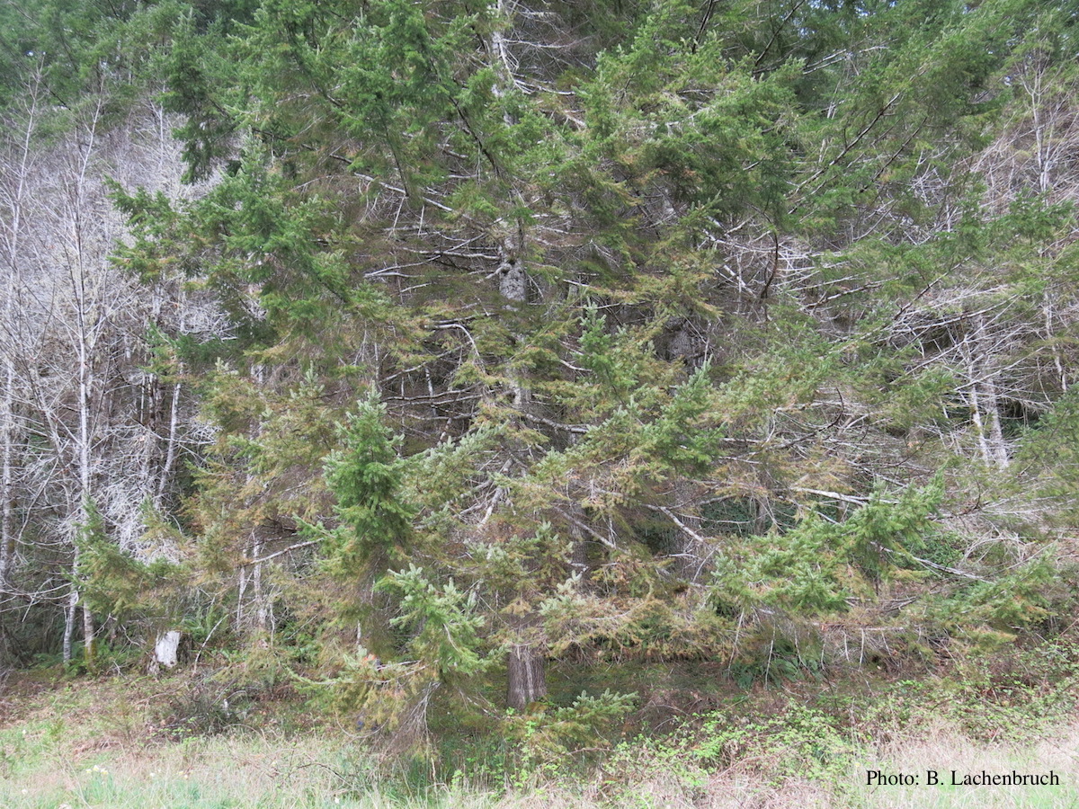 Red needle cast symptoms on Douglas-fir in western Oregon, 2015 |
|
P. cinnamomi on Fraser fir 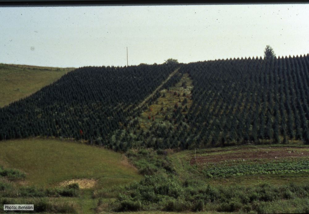 Frasier fir Christmas trees, North Carolina |
P. cactorum bleeding canker 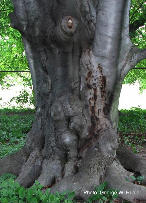 Bleeding canker on European beech (Fagus sylvatica) |
P. cambivora bole canker 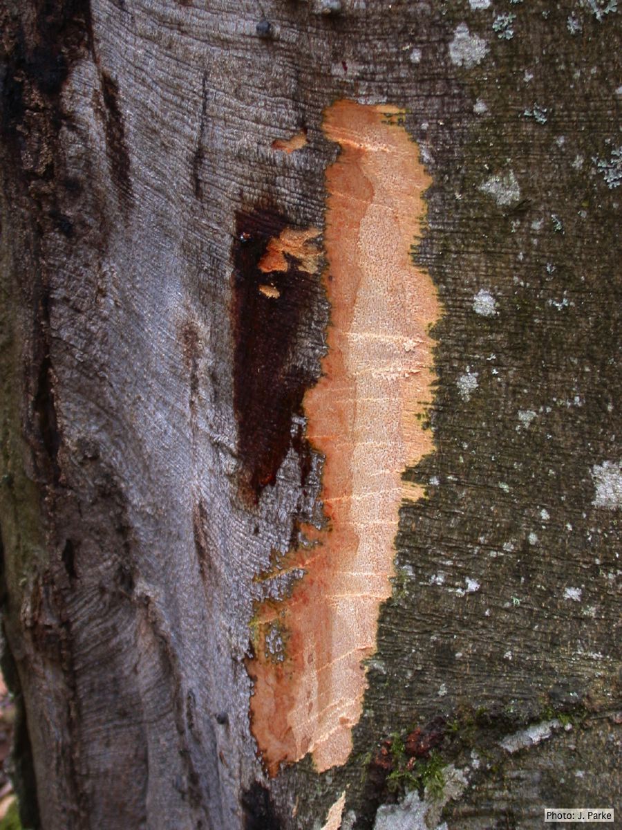 Fagus sylvatica bole canker |
|
P. alni in riparian alder, Germany 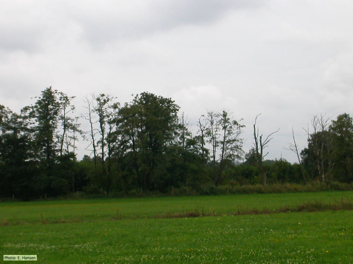 P. alni in riparian alder, Germany |
P. cactorum sporangium  P. cactorum sporangium |
P. agathidicia disease symptoms on kauri 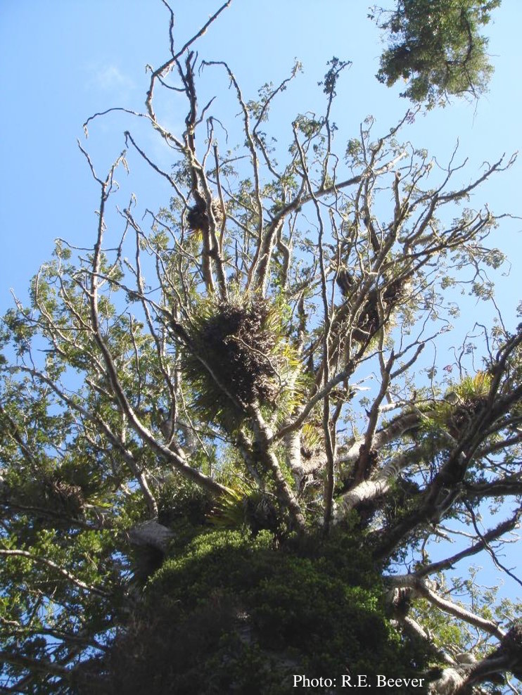 Crown decline of mature kauri, with branchlets with little or no leaves |