

Disease symptoms on a cocoa pod
Photo Gallery
Site will be retired 9/1/2026
This site is no longer being developed and will be retired on September 1, 2026. Please contact us if you have any questions or would like to provide support to continue the project.
|
P. megakarya disease symptoms on Theobroma cacao fruit 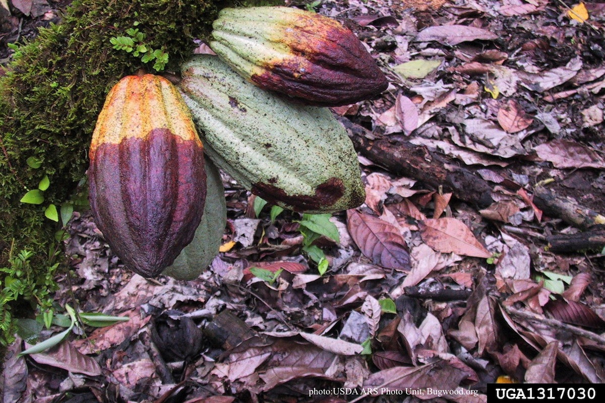 |
P. cambivora coralloid irregular hyphae  Coralloid irregular hyphae |
P. nemorosa colony morphology on PDA  Colony morphology on PDA at 14 days |
|
P. austrocedrae - Mal del ciprés, stages of decline 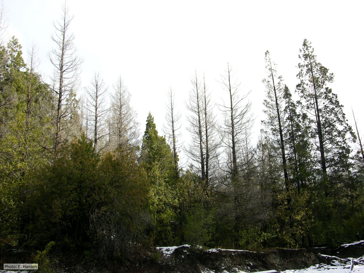 Colony morphology of P. austrocedrae at 16 C after four weeks on PDA |
P. lateralis on Port Orford Cedar 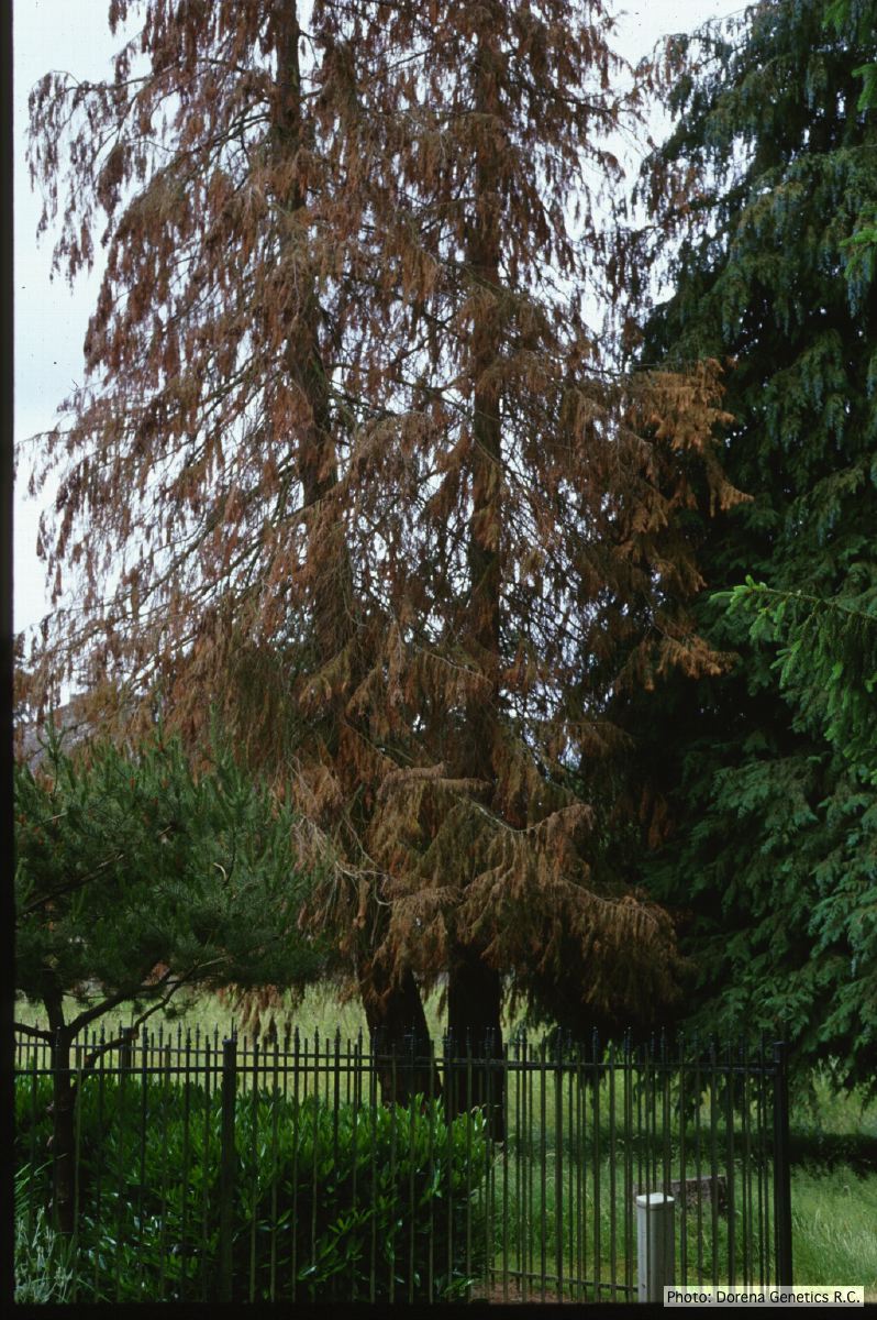 Horticultural planting of Port-Orford cedar, photo from USDA Forest Service, Dorena Genetic Resource Center |
P. katsurae oogonia 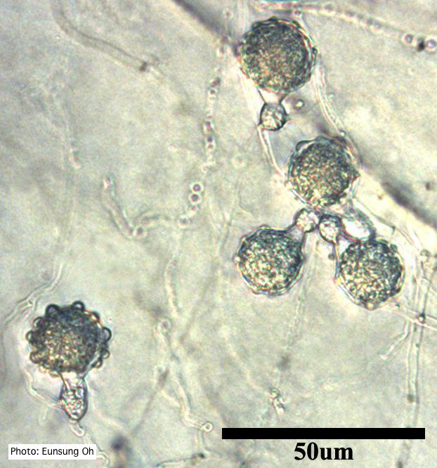 Oogonia with ornamentation |
|
P. kernoviae sporangium 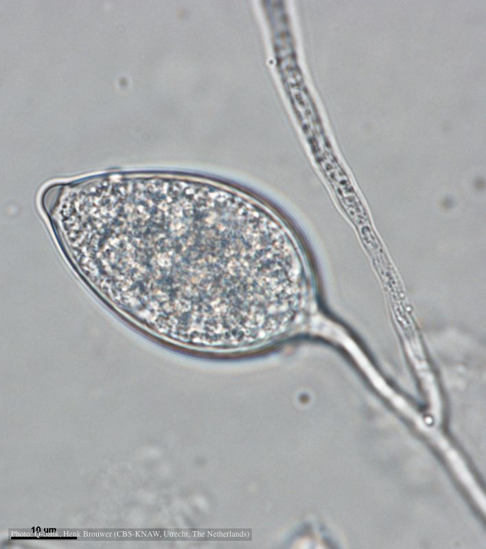 Papillate and caducous sporangium, photo from Q-bank, used with permission |
P. palmivora sporangia 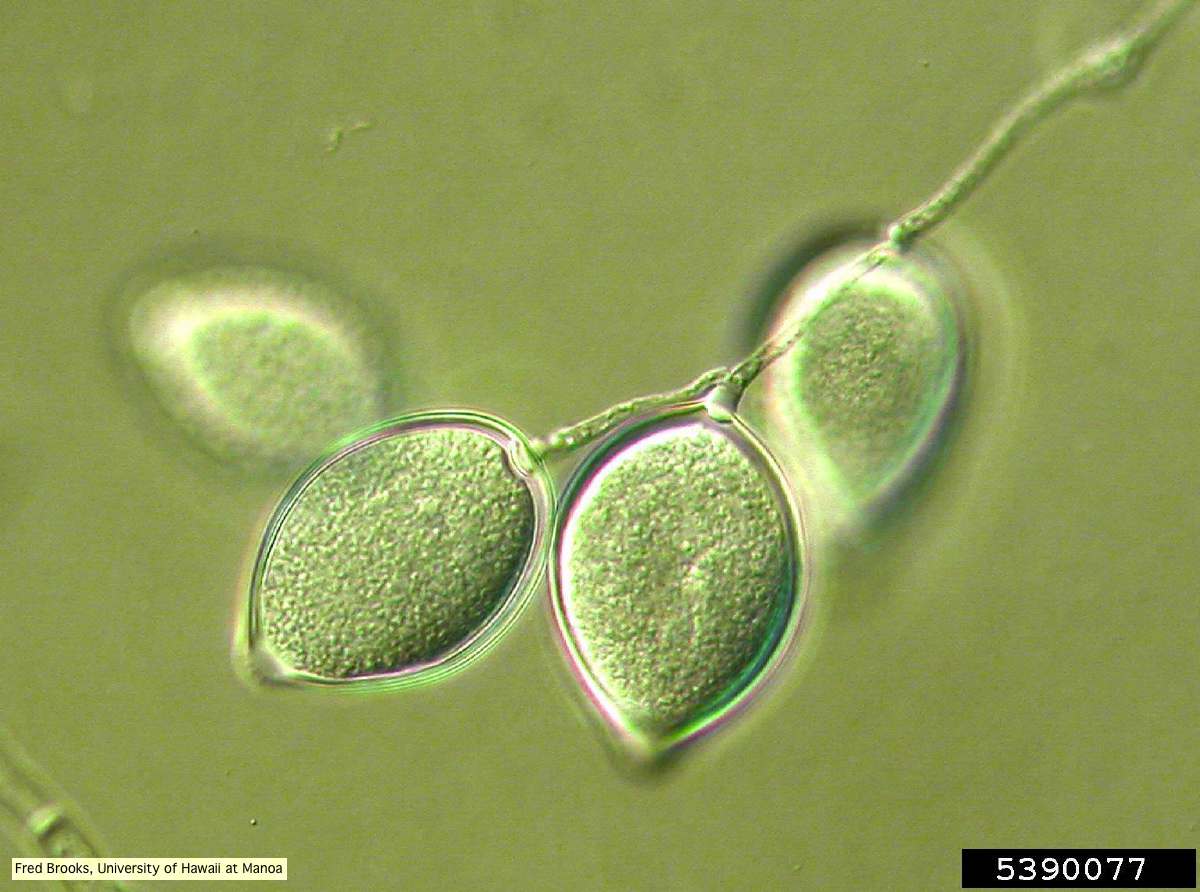 Sporangia (sporangiospores) showing sympodial branching |
P. pinifolia on Pinus radiata  Pinus radiata, note Infected needles at right angles to stem |
|
P. tentaculata sporangium 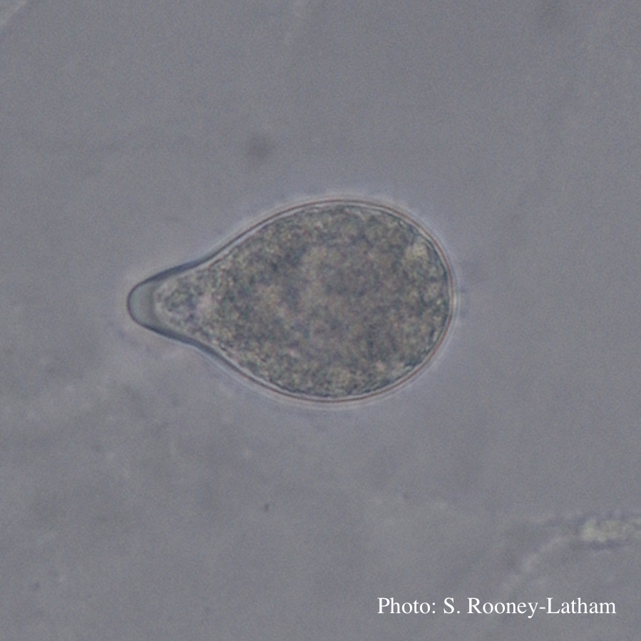 Papillate sporangium of P. tentaculata with an elongated neck or beak. |
Dying Port Orford Cedar trees 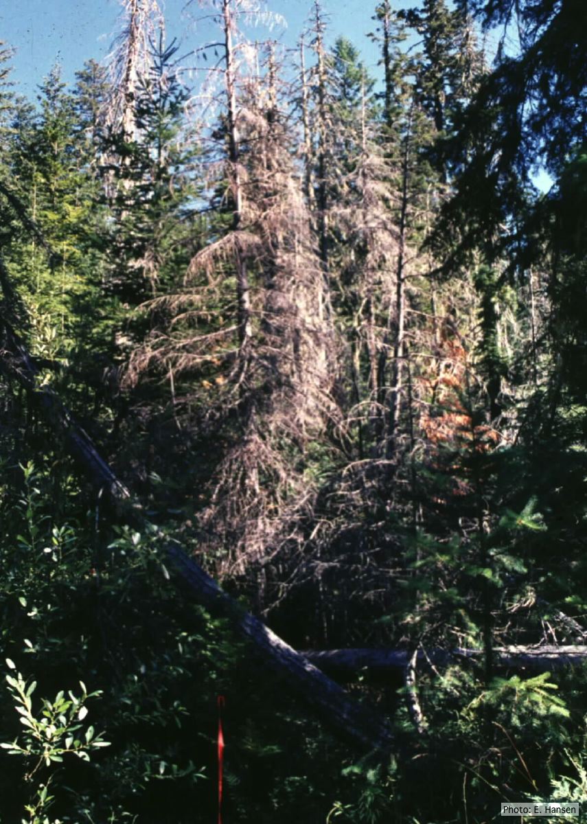 Dead Chamaecyparis lawsoniana trees |
P. cambivora sporangium 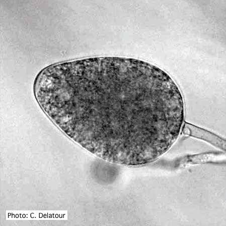 Ovoid non- papillate sporangia |