

Dieback of Juglans regia caused by Phytophthora cactorum
Photo Gallery
Site will be retired 9/1/2026
This site is no longer being developed and will be retired on September 1, 2026. Please contact us if you have any questions or would like to provide support to continue the project.
|
Phytophthora cactorum disease symptoms on English walnut  |
P. cambivora colony morphology on PDA 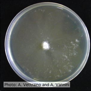 Appressed colony morphology at 14 days at 20°C on PDA |
P. cambivora disease symptoms 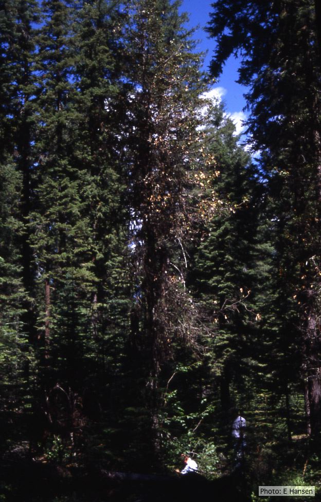 Dead and dying chinquapin infected with P. cambivora |
|
P. ramorum sporangium  Deciduous sporangium, photo from Q-bank, used with permission |
P. nicotianae colony morphology on PDA 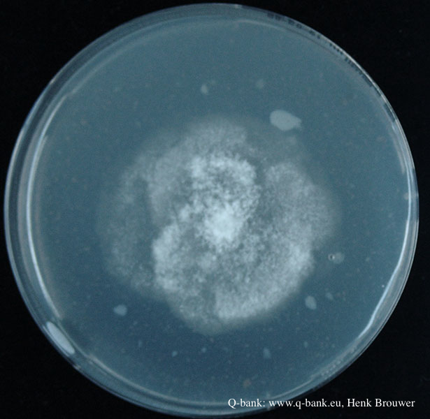 Phytophthora nicotianae CBS 321.49 PDA after 7 days at 24 degrees. Photo from Q-bank: www.q-bank.eu, Henk Brouwer (CBS-KNAW, Utrecht, The Netherlands) |
P. pluvialis on Pinus radiata in New Zealand 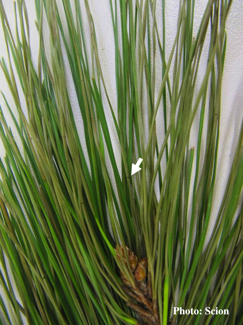 A Pinus radiata needle showing faded olive- or khaki- coloured lesions consistent with the presence of red needle cast disease. Arrow shows resinous bands within the extended olive lesion. |
|
P. pluvialis symptoms on Douglas-fir needles 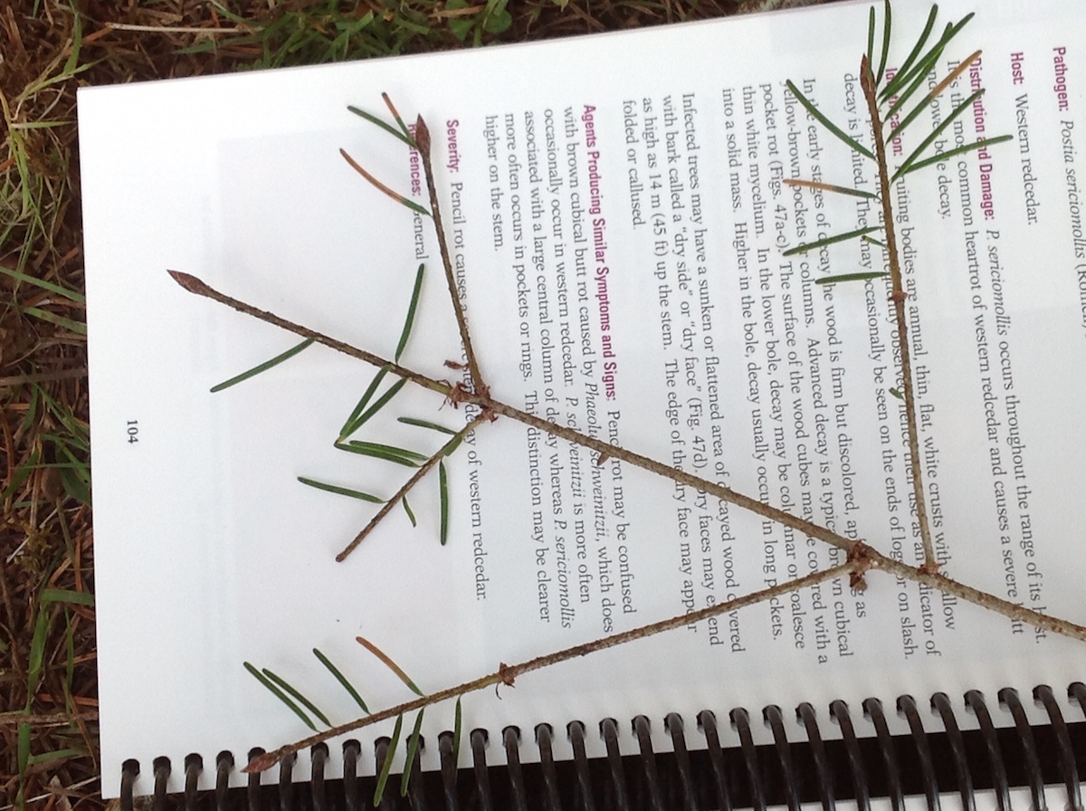 Symptoms of red needle cast on Douglas-fir needles |
P. pseudosyringae sporangium 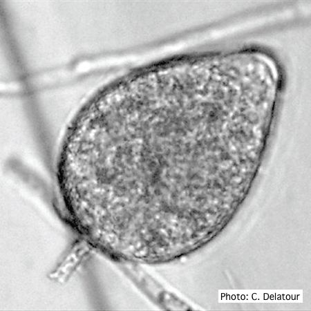 Ovoid, semipapillate sporangia showing medium length pedicel |
P. cambivora oogonium 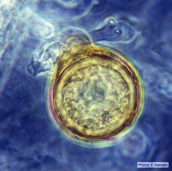 P. cambivora oogonium with antheridium |
|
Growth morphology on V8 of P. lateralis 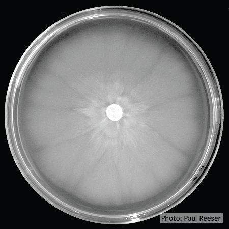 Colony morphology on V8 at 14 days |
P. ramorum colony morphology on CMA PARP 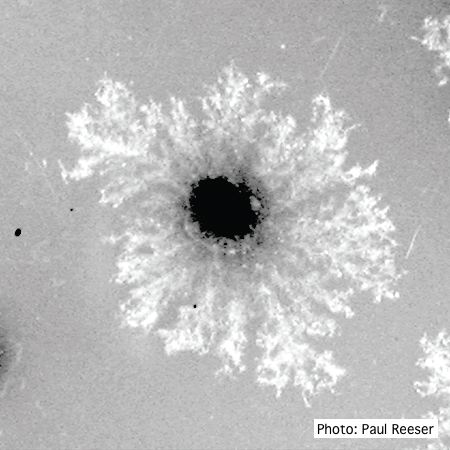 P. ramorum colony morphology on CMA PARP |
P. tentaculata oogonia and antheridia 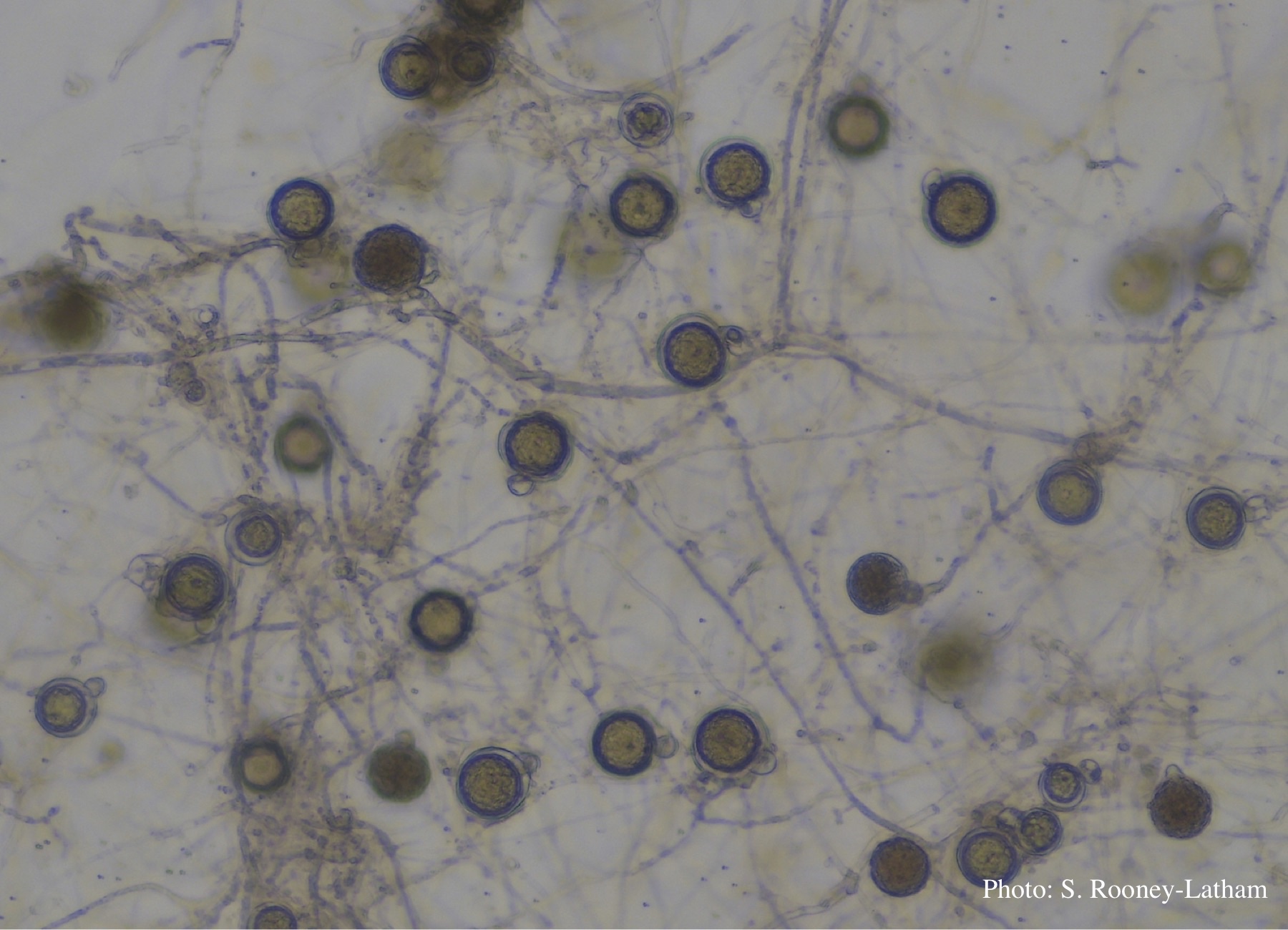 Oospores and oogonia with mostly paragynous but some amphigynous antheridia of P. tentaculata |