

Kauri tree in New Zealand with disease symptoms
Photo Gallery
Site will be retired 9/1/2026
This site is no longer being developed and will be retired on September 1, 2026. Please contact us if you have any questions or would like to provide support to continue the project.
|
Phytophthora taxon Agathis bole canker  |
P. austrocedrae - sporangia 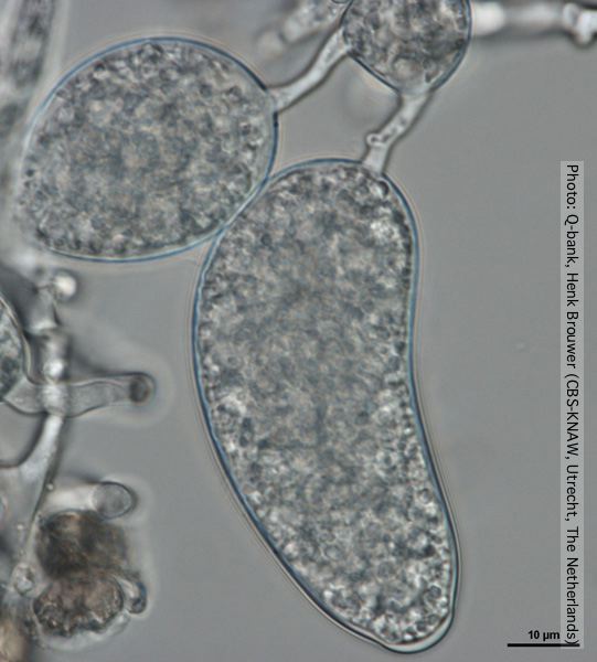 Sporangium with distorted shape, photo from Q-bank, used with permission. |
Growth of P. megakarya on V8 agar 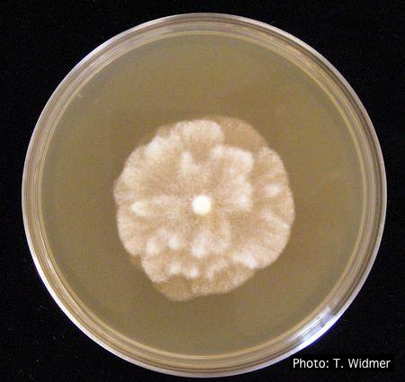 Growth of P. megakarya on V8 agar |
|
P. cactorum bleeding canker  Bleeding canker on European beech (Fagus sylvatica) |
P. nemorosa colony morphology on PDA  Colony morphology on PDA at 14 days |
P. cryptogea colony morpholgy on V8  Colony morphology on V8 at 14 days |
|
P. pseudosyringae hyphal swellings  Sub-globose hyphal swellings in water |
P. nicotianae symptoms 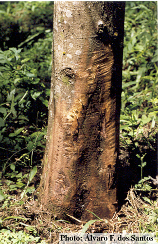 Symptoms of gummosis on black wattle (Fitopatol. bras. 2005) |
P. austrocedrae oogonium 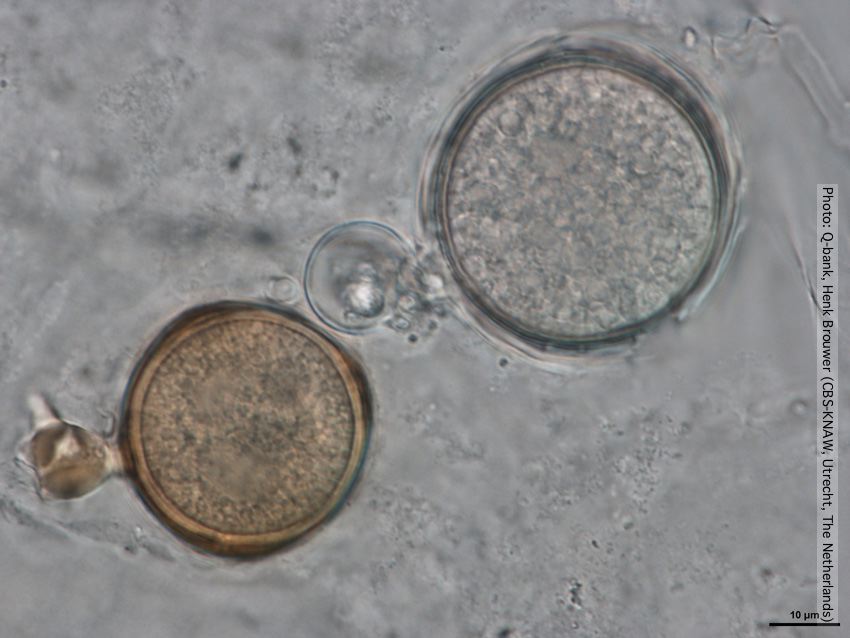 Oogonia with and without brown pigment, photo from Q-bank, used with permission |
|
P. ramorum colony morphology on V8 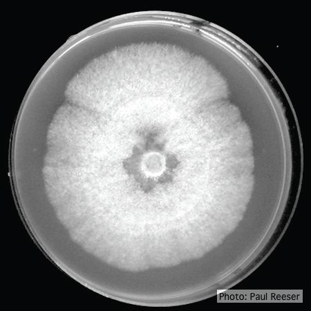 P. ramorum colony morphology on V8 |
Phytophthora cactorum disease symptoms on English walnut  Dieback of Juglans regia caused by Phytophthora cactorum |
Tāne Mahuta “Lord of the Forest” kauri tree 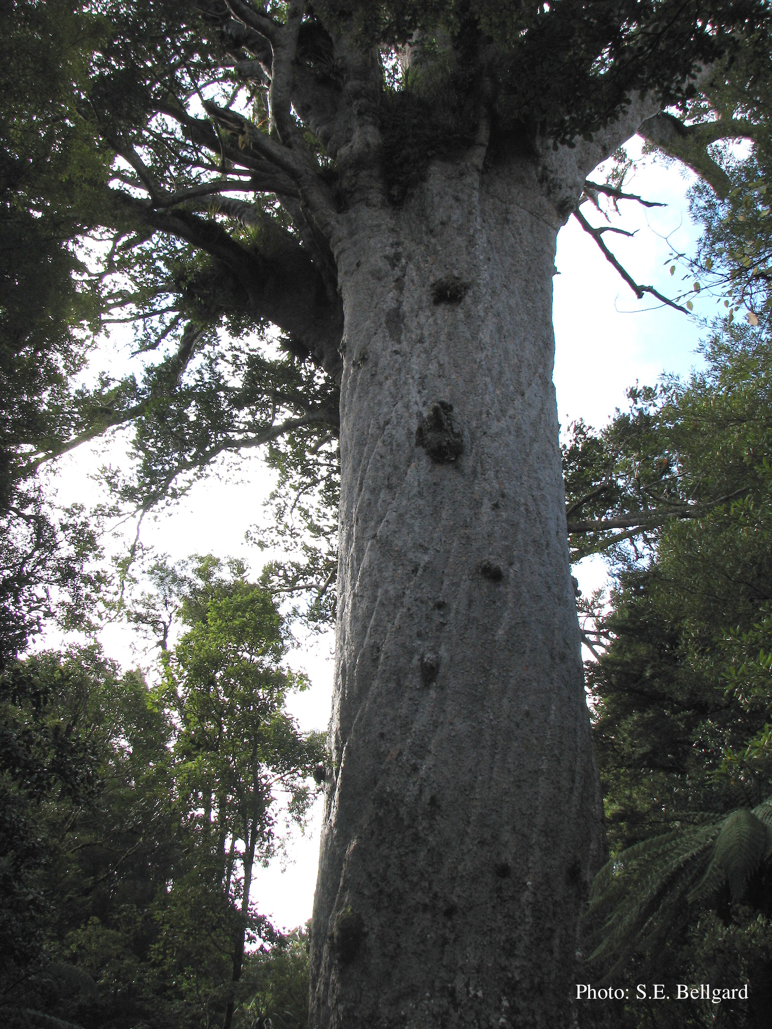 Tāne Mahuta “Lord of the Forest” is a giant kauri tree (approximately 47 metres in height) in the Waipoua Forest of Northland Region, New Zealand. Its age is unknown but is estimated to be between 1,250 and 2,500 years |