

Phytophthora chlamydospora sporangium in water. Bar is 20µm.
Photo Gallery
Site will be retired 9/1/2026
This site is no longer being developed and will be retired on September 1, 2026. Please contact us if you have any questions or would like to provide support to continue the project.
|
P. chlamydospora sporangium  |
P. lateralis on Port Orford Cedar 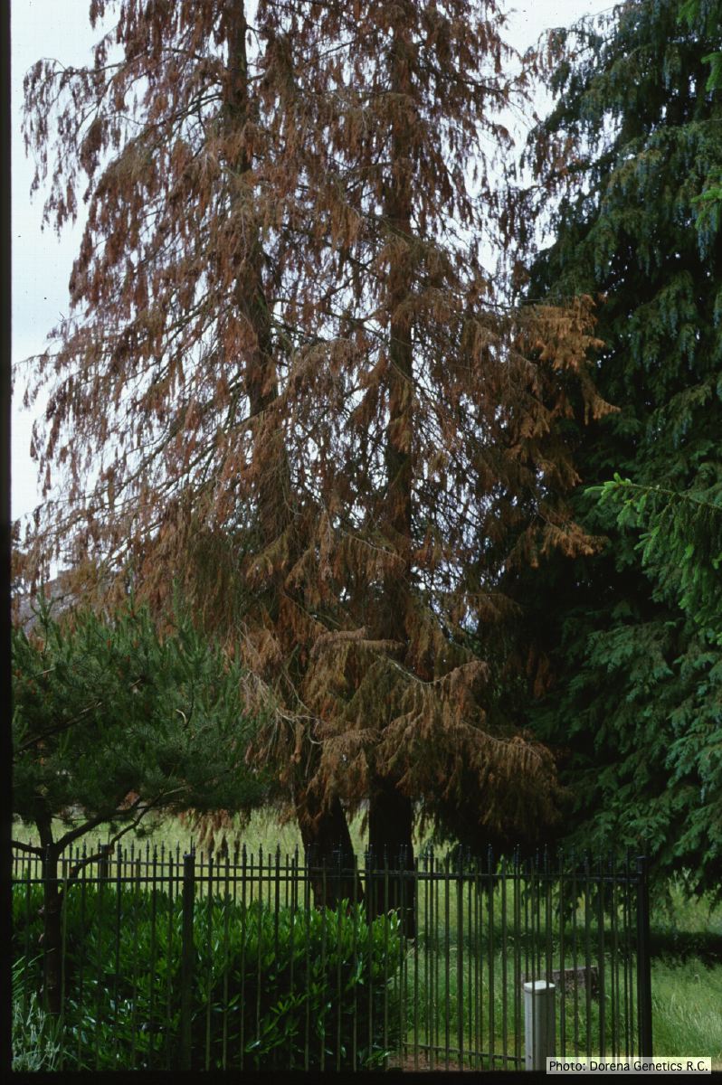 Horticultural planting of Port-Orford cedar, photo from USDA Forest Service, Dorena Genetic Resource Center |
P. boehmeriae oogonia  Oogonia and oospores with amphigynous antheridia |
|
P. austrocedrae colony morphology on Tomato juice agar 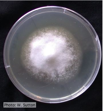 Colony morphology of P. austrocedrae at 16 ºC after 4 weeks on Tomato juice agar |
P. cinnamomi colony morphology on PDA  P. cinnamomi colony growth on PDA at 14 days |
P. tentaculata chlamydospore 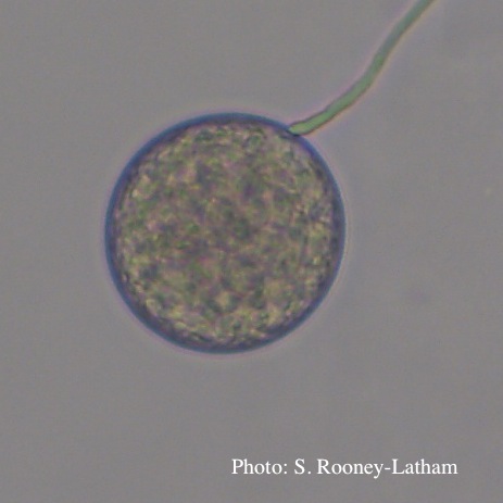 Terminal chlamydospore of P. tentaculata |
|
P. austrocedrae - Mal del ciprés, stages of decline 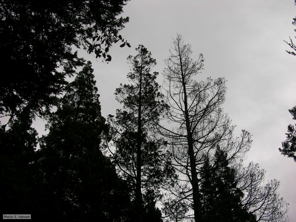 Mal del ciprés, stages of decline |
Chlamydospore of P. lateralis 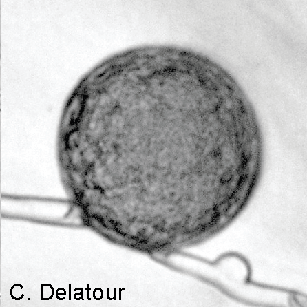 Laterally intercalary chlamydospore of Phytophthora lateralis |
P. cambivora sporangium with internal extended proliferation  Empty sporagia showing internal nested and extended proliferation |
|
Growth of P. arenaria on CA  Colony morphology of Phytophthora arenaria after 7 days at 20°C on carrot agar |
P. cryptogea colony morpholgy on V8  Colony morphology on V8 at 14 days |
P. pinifolia sporulation 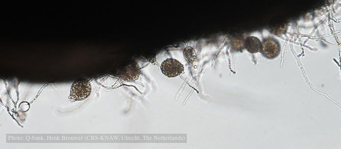 Sporulation on edge of hemp seed, photo from Q-bank, used with permission. |