

Inactive lesion of P. cambivora on chinquapin
Photo Gallery
Site will be retired 9/1/2026
This site is no longer being developed and will be retired on September 1, 2026. Please contact us if you have any questions or would like to provide support to continue the project.
|
P. cambivora inactive lesion on chinquapin  |
P. alni oogonium 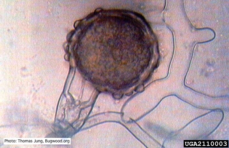 Bullate oogonium of P. alni (German variant) with oospore and amphigynous antheridium. |
P. cambivora symptoms 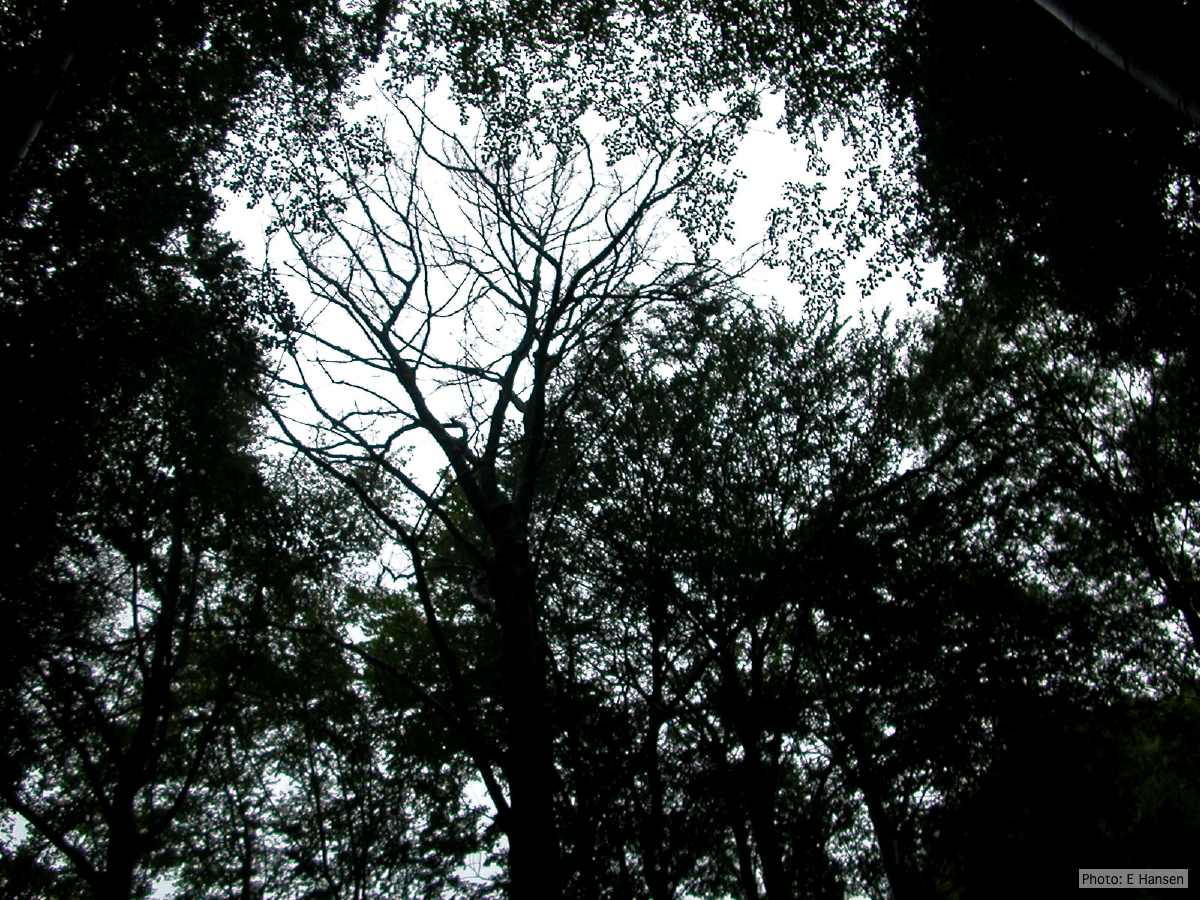 Dead beech in Germany |
|
Dead and healthy Port-Orford cedar seedlings  Port-Orford-cedar seedlings planted to test for Phytophthora lateralis resistance at the Dorena Genetic Resource Center |
P. tentaculata hyphae 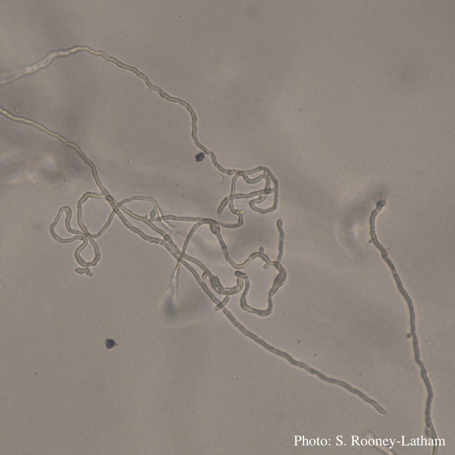 Looping hyphae commonly seen with P. tentaculata on PARP media |
P. cambivora colony morphology on PDA  Rosaceous colony morphology at 14 days at 20°C on PDA |
|
P. cambivora colony morphology on PDA 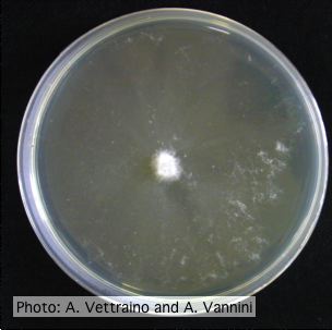 Appressed colony morphology at 14 days at 20°C on PDA |
P. kernoviae sporangium 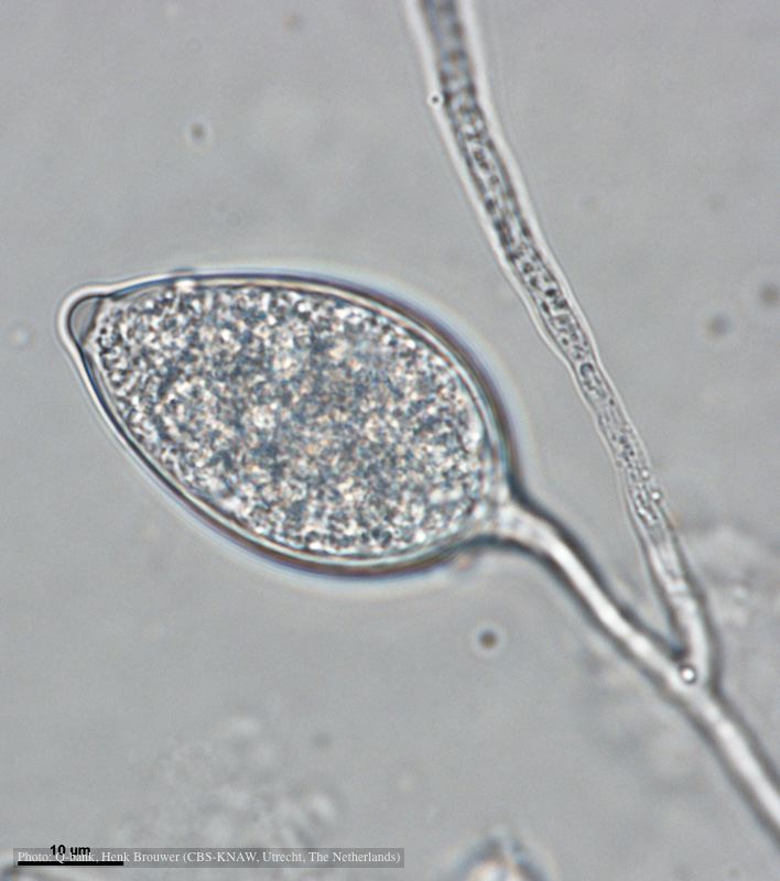 Papillate and caducous sporangium, photo from Q-bank, used with permission |
P. cactorum oogonium 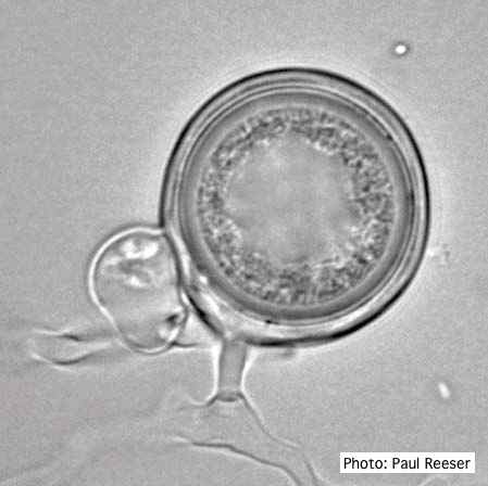 Oogonium with paragynous antheridia close to oogonial stalk. Oospores are slightly aplerotic. |
|
P. pinifolia on Pinus radiata 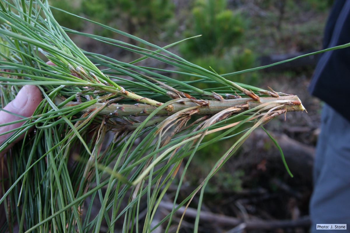 Pinus radiata, note grey and collapsed needle bases |
P. pseudosyringae oogonium  Oogonium with paragynous antheridium in agar |
Austrocedrus associated with Mal del ciprés.  Austrocedrus with basal resin flow associated with Mal del ciprés. |