

P. alni in alder forest, Germany, with T. Jung
Photo Gallery
Site will be retired 9/1/2026
This site is no longer being developed and will be retired on September 1, 2026. Please contact us if you have any questions or would like to provide support to continue the project.
|
P. alni in alder forest, Germany, with T. Jung 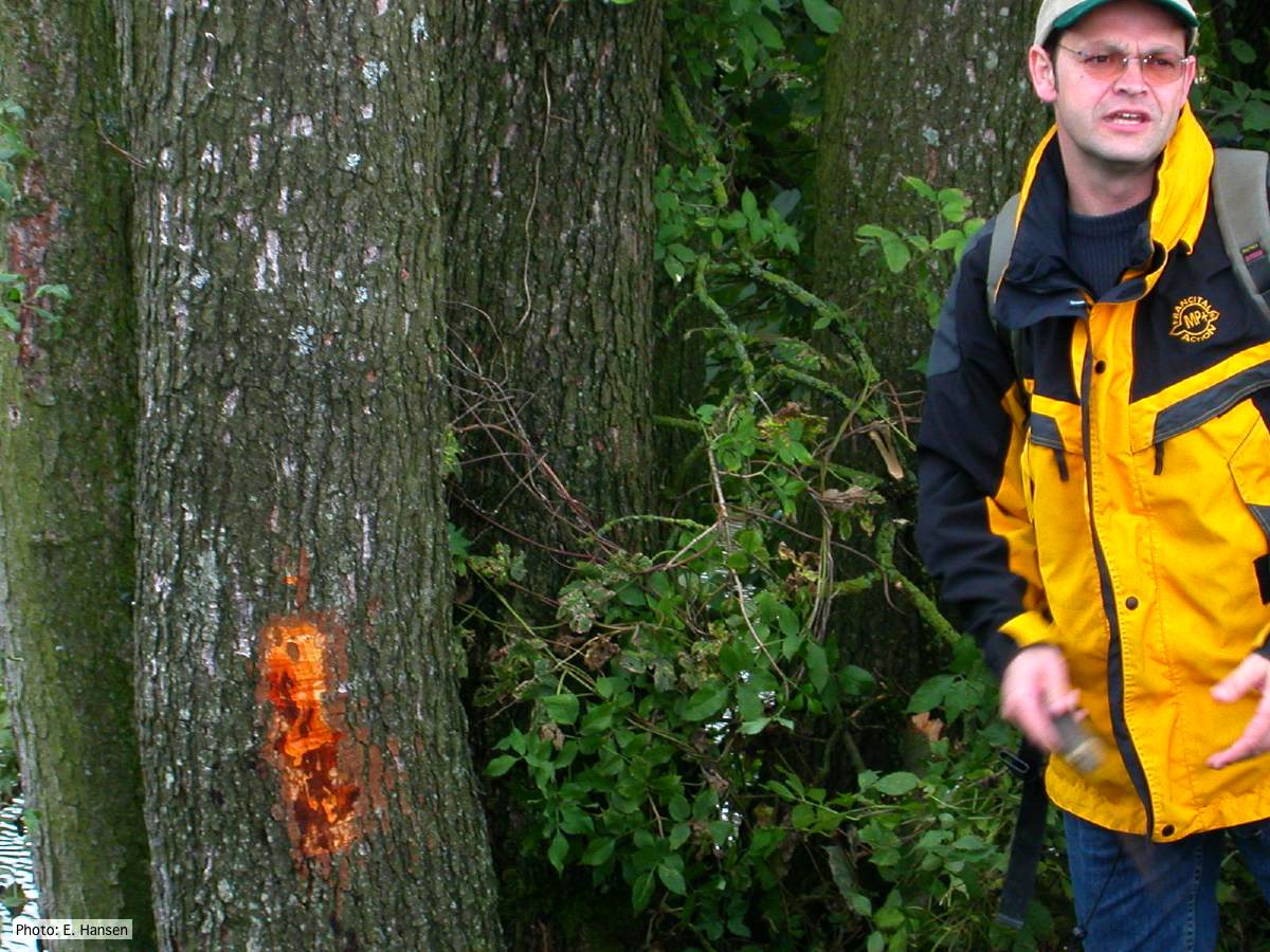 |
P. siskiyouensis canker on Italian alder 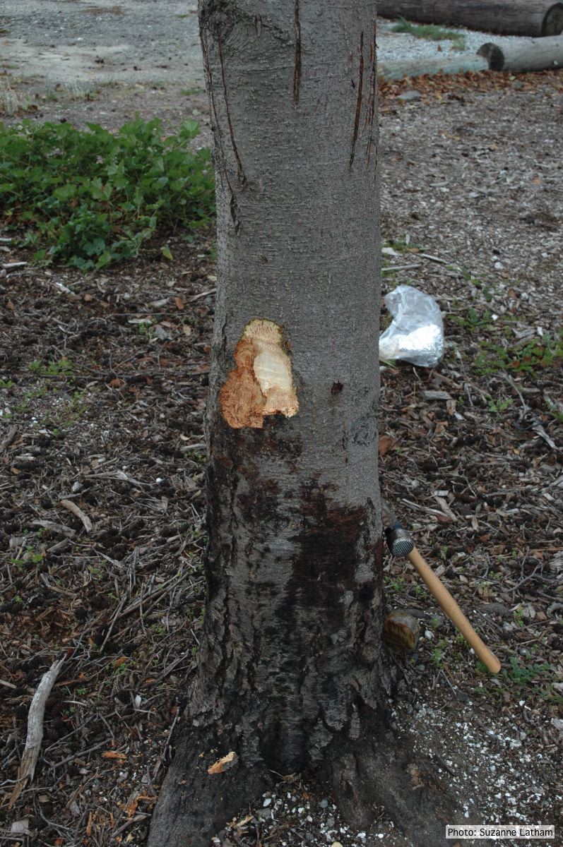 Bole lesions in the tissues under the bark of a bleeding canker: distinct margin between healthy and disease tissues |
P. pluvialis on Pinus radiata in New Zealand  Typical red needle cast symptoms along a twig. Lesions begin at the base of the needle which subsequently turns brown and is cast from the twig. |
|
P. cambivora colony morphology on MA 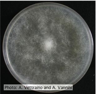 Uniform fluffy colony morphology at 14 days at 20°C on MA |
P. austrocedrae - Mal del ciprés, stages of decline 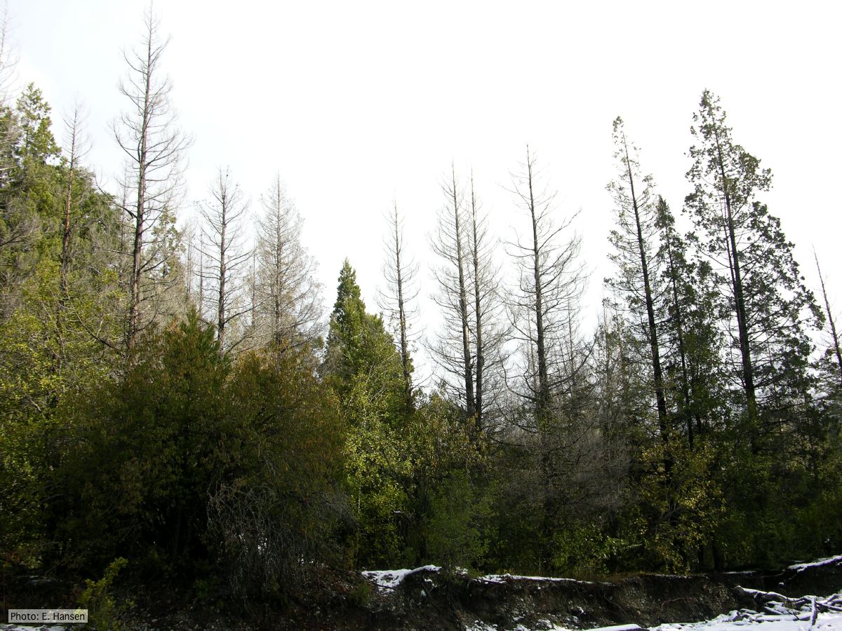 Colony morphology of P. austrocedrae at 16 C after four weeks on PDA |
P. nicotianae sporangia  P. nicotianae overview of sporangia 40x. Photo from Q-bank: www.q-bank.eu, Henk Brouwer (CBS-KNAW, Utrecht, The Netherlands) |
|
P. nemorosa colony morphology on PDA  Colony morphology on PDA at 14 days |
P. austrocedrae hyphal swellings in liquid media drawing  Morphology of hyphae of Phytophthora austrocedrae, from Greslebin et al. 2007 |
P. tentaculata oogonia and antheridia 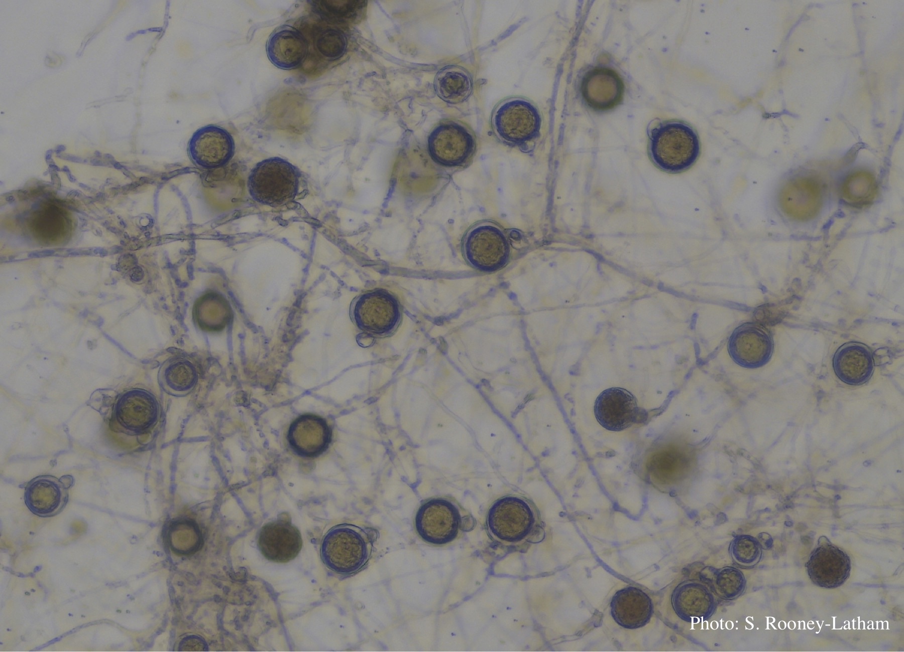 Oospores and oogonia with mostly paragynous but some amphigynous antheridia of P. tentaculata |
|
P. lateralis on Port Orford Cedar 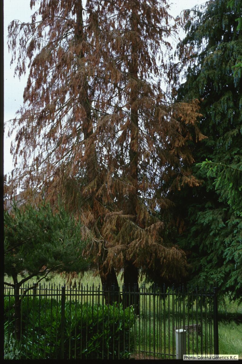 Horticultural planting of Port-Orford cedar, photo from USDA Forest Service, Dorena Genetic Resource Center |
P. lateralis on Port Orford cedar  Localized branch infection of Chaemacyparis lawsoniana in Lopérec, France |
P. austrocedrae colony morphology on Tomato juice agar with B sitosterol 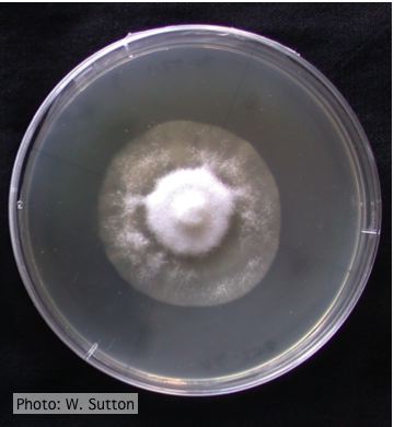 Colony morphology of P. austrocedrae at 16 ºC after 4 weeks on Tomato juice agar with B sitosterol |