

Colony morphology on V8 at 14 days
Photo Gallery
Site will be retired 9/1/2026
This site is no longer being developed and will be retired on September 1, 2026. Please contact us if you have any questions or would like to provide support to continue the project.
|
P. cambivora colony morphology on V8 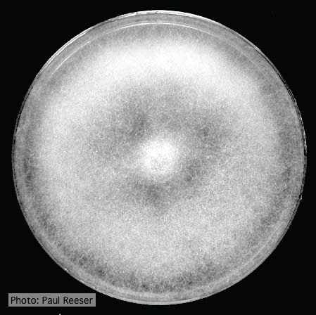 |
P. palmivora colony morphology on V8 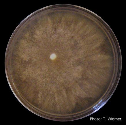 Growth of P. palmivora on V8 agar |
P. nemorosa oogonium 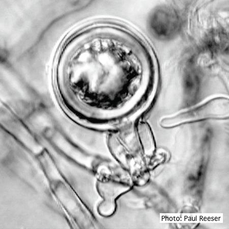 Oogonium with amphigynous antheridium |
|
P. arenaria disease symptoms on Banksia landscape 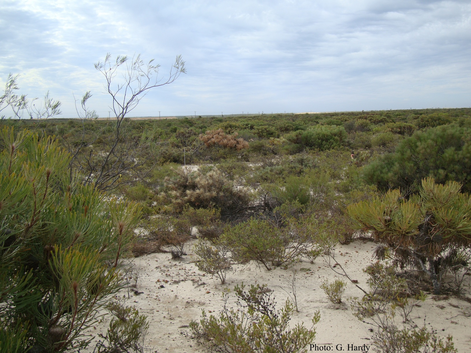 Dead Banksia sp. in a Kwongan heathland on mineral sand near Eneabba, Western Australia recently killed by root and collar rot caused by Phytophthora arenaria |
P. tentaculata sporangium 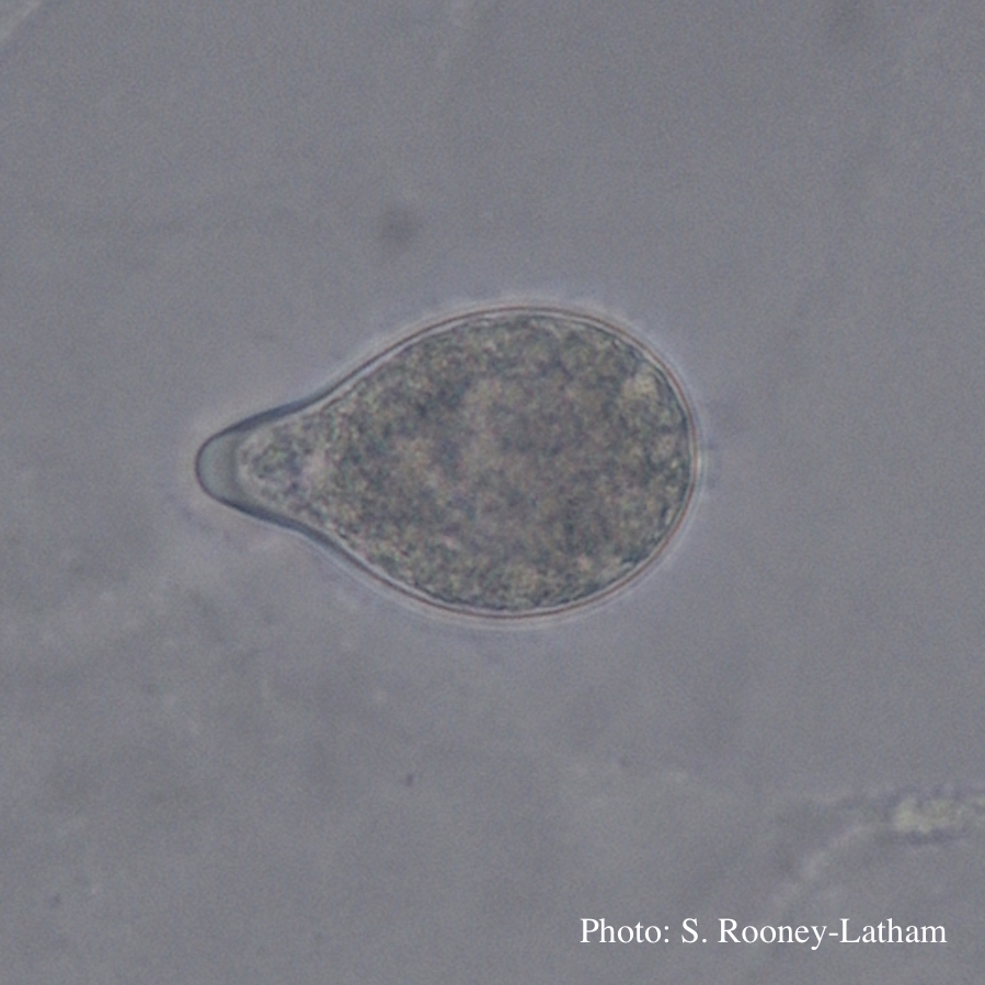 Papillate sporangium of P. tentaculata with an elongated neck or beak. |
P. lateralis on Port Orford cedar  Atypical decline caused by aerial infections in Scaër, France |
|
Dying Port Orford Cedar trees 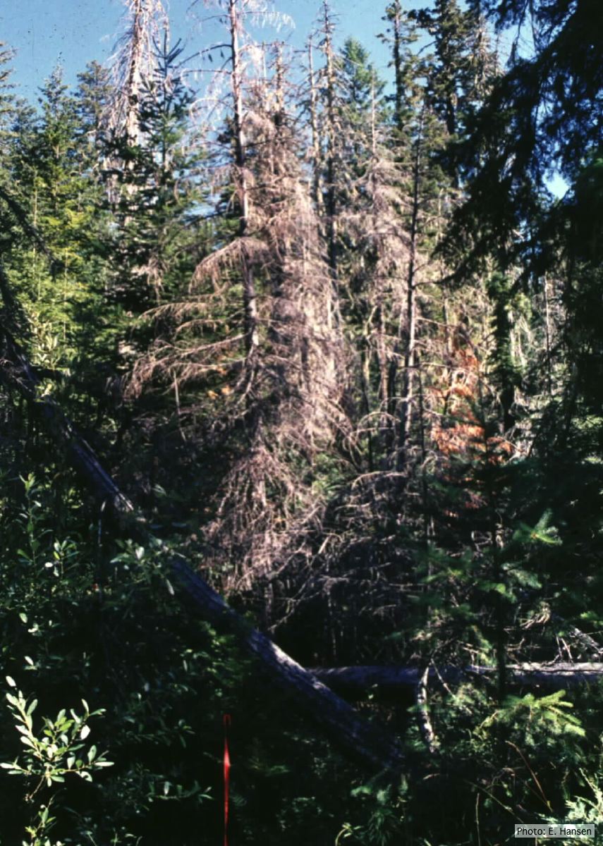 Dead Chamaecyparis lawsoniana trees |
P. cambivora disease symptoms 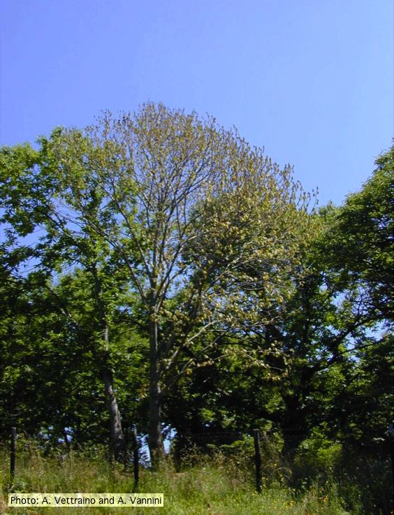 Crown symptoms of Ink disease on sweet chestnut |
P. austrocedrae - necrotic lesion in phloem 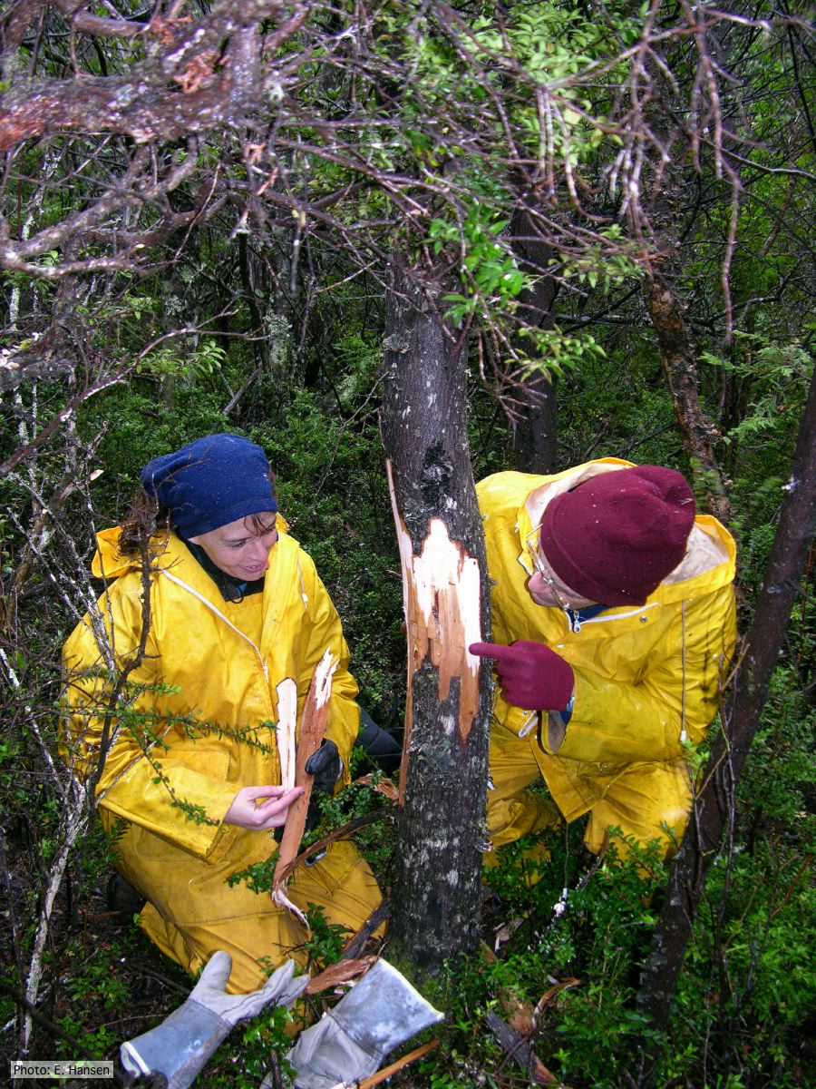 |
|
P. agathidicia growth on PDA 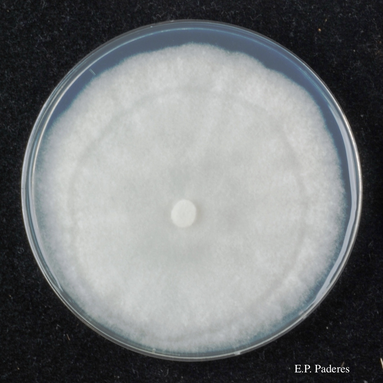 Colony morphology of ex-holotype ICMP 17027 after 10-days incubation at 20°C in the dark |
P. chlamydospora hyphal swellings 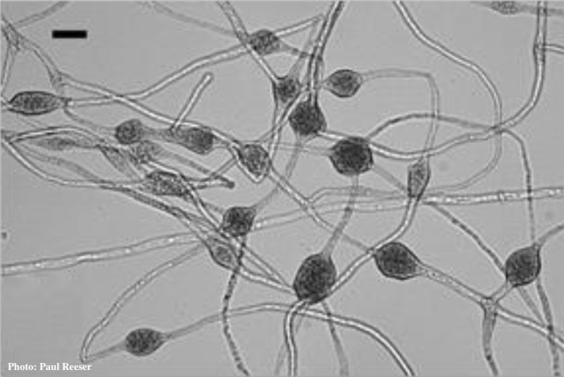 Phytophthora chlamydospora chlamydospore in agar. Bar is 20µm.
|
P. lateralis on Port Orford cedar 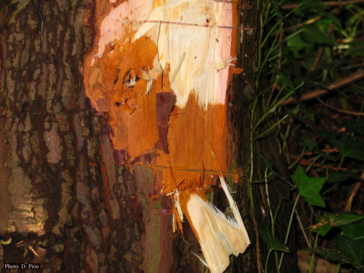 Collar lesion on Chaemacyparis lawsoniana in Landrévarzec, France |