

Colony morphology on PDA at 14 days
Photo Gallery
Site will be retired 9/1/2026
This site is no longer being developed and will be retired on September 1, 2026. Please contact us if you have any questions or would like to provide support to continue the project.
|
Basal canker on Port Orford Cedar stump  |
P. cryptogea colony morpholgy on PDA 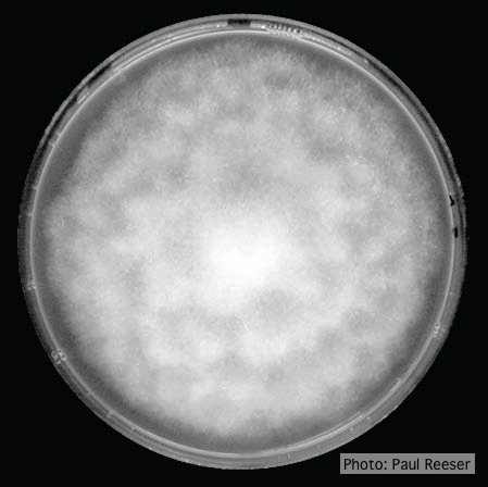 |
P. siskiyouensis disease symptoms on Italian alder 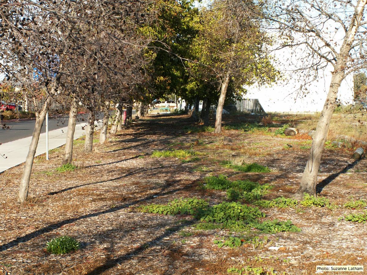 Grove of dying trees in a commercial landscape in Foster City, CA |
|
Dead and healthy Port-Orford cedar seedlings  Port-Orford-cedar seedlings planted to test for Phytophthora lateralis resistance at the Dorena Genetic Resource Center |
P. cinnamomi cork oak decline  Cork oak decline, Portugal |
P. katsurae sporangia 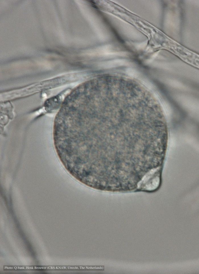 Papillate, non-caducous sporangium with differentiated content; photo used with permission from Q-bank |
|
P. chlamydospora sporangium 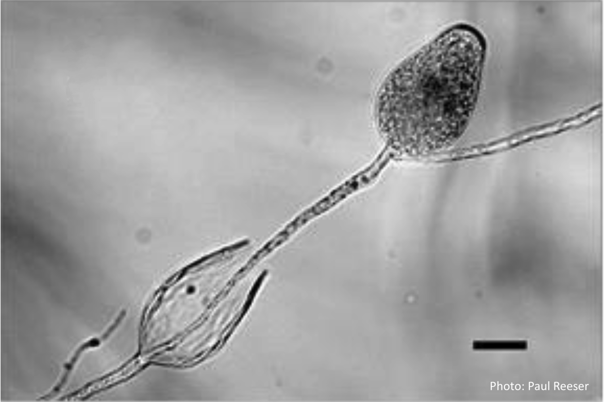 Phytophthora chlamydospora sporangia in water, showing subsporangial elongation. Bar is 20 µm. |
P. austrocedrae colony morphology on PDA  Colony morphology of P. austrocedrae at 16 C after four weeks on PDA |
P. pinifolia on Pinus radiata  Pinus radiata, note Infected needles at right angles to stem |
|
P. cambivora colony morphology on MA  Appressed colony morphology at 14 days at 20°C on MA |
P. cryptogea sporangium  Obpyriform non-papillate sporangia in water |
P. pinifolia on Pinus radiata 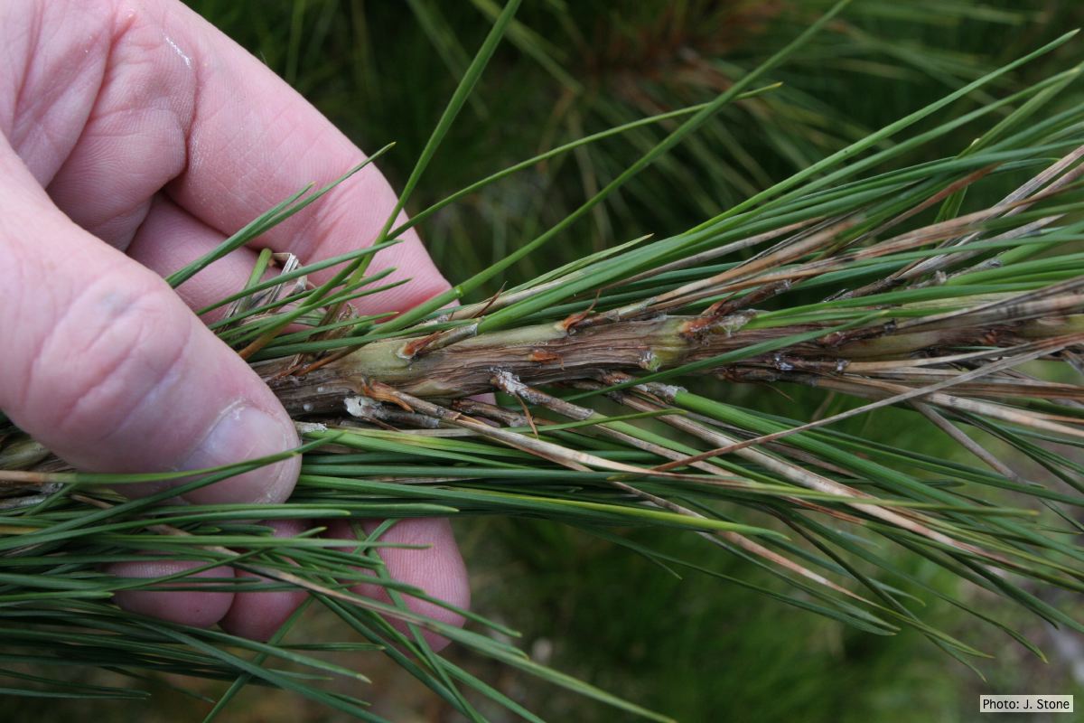 Pinus radiata needles, note “black line” symptom near needle bases |