

Noncaducous sporangium showing ovoid shape and papillate condition. (Fitopatol. bras. 2005)
Photo Gallery
Site will be retired 9/1/2026
This site is no longer being developed and will be retired on September 1, 2026. Please contact us if you have any questions or would like to provide support to continue the project.
|
P. nicotianae sporangia  |
P. kernoviae disease on beech 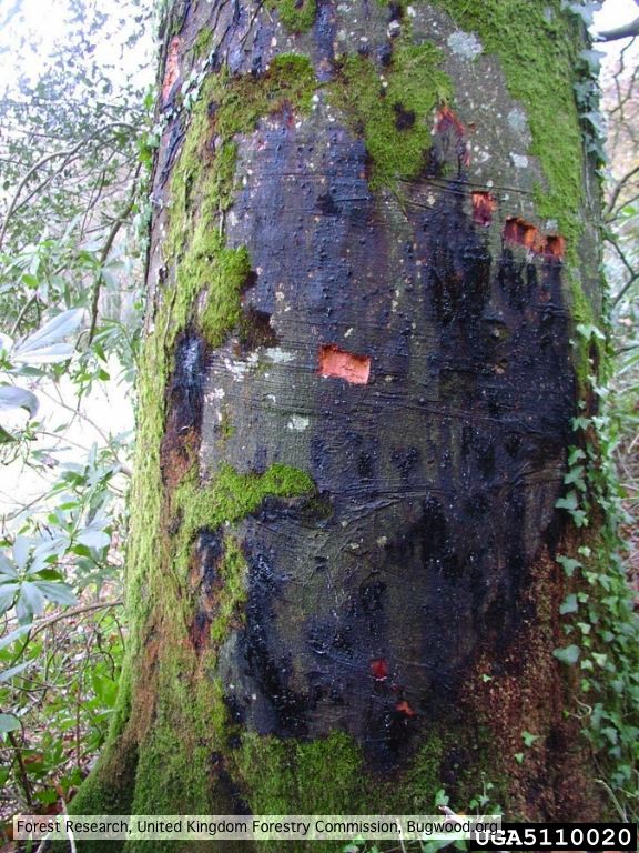 External lesion; 14 November 2003 |
P. austrocedrae oogonium 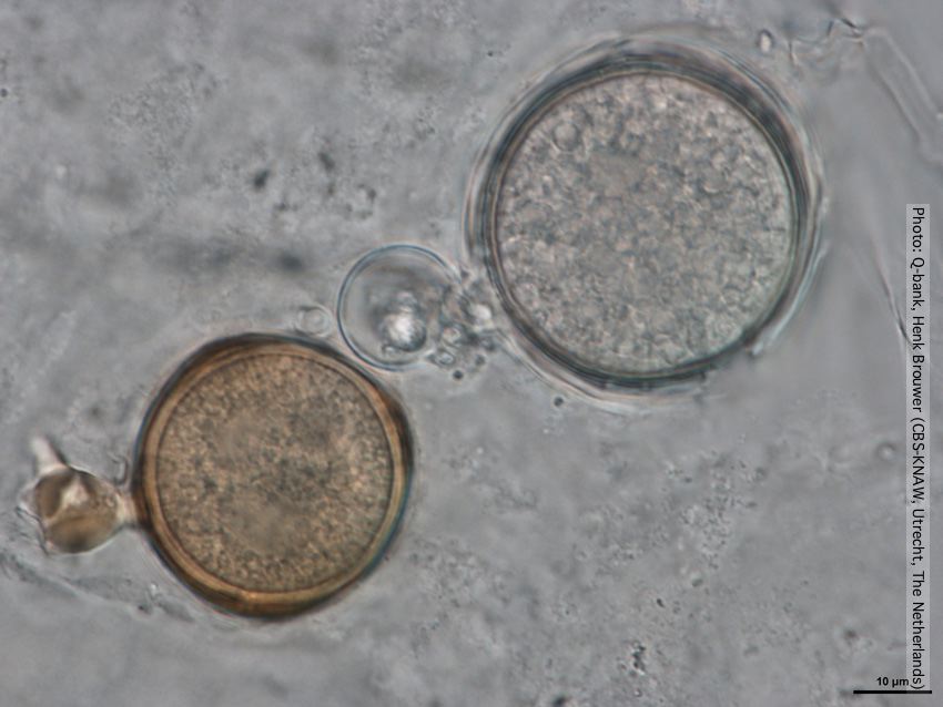 Oogonia with and without brown pigment, photo from Q-bank, used with permission |
|
P. agathidicida lesion on kauri tree 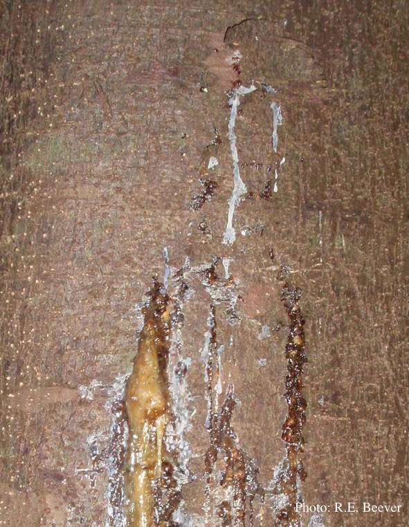 Gum oozing out of longitudinal lesion |
P. austrocedrae - Mal del ciprés, stages of decline 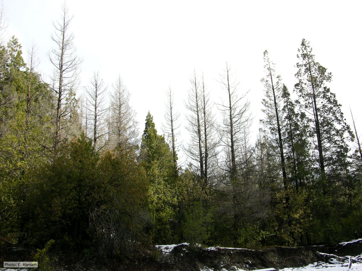 Colony morphology of P. austrocedrae at 16 C after four weeks on PDA |
Black pod disease of cacao 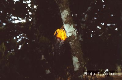 Symptom of black pod disease of cacao (T. cacao) caused by P. palmivora |
|
P. pseudosyringae hyphal swellings  Sub-globose hyphal swellings in water |
P. cinnamomi on Banksia 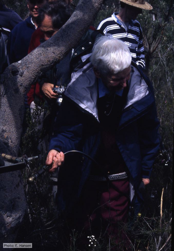 Gretna Weste injecting Banksia with phosphonate |
P. cambivora colony morphology on PDA 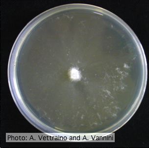 Appressed colony morphology at 14 days at 20°C on PDA |
|
P. tentaculata disease symptoms on California mugwort 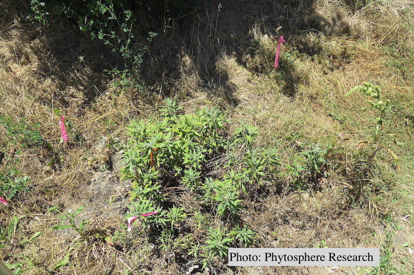 Outplanted California mugwort (Artemisia douglasiana) infected with P. tentaculata, 4.5 years after planting. Plant shows stunting and chlorosis. (P. cryptogea and P. lacustris were also baited from roots/soil of this plant). |
P. tentaculata chlamydospore  P. tentaculata chlamydospore with short hyphal projection |
P. alni sporangia 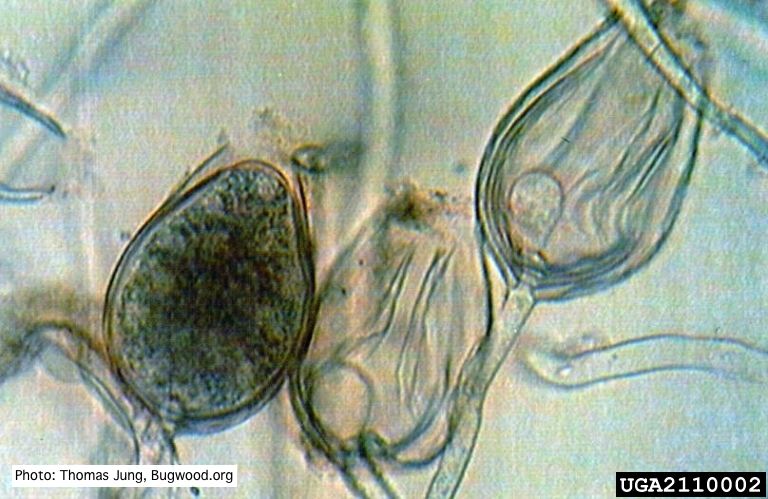 Non-papillate sporangia of P. alni showing nested proliferation. |