

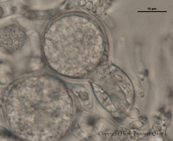
‘Blistered’ hyphal swellings in agar
Photo Gallery
Site will be retired 9/1/2026
This site is no longer being developed and will be retired on September 1, 2026. Please contact us if you have any questions or would like to provide support to continue the project.
|
P. nemorosa hyphal swellings 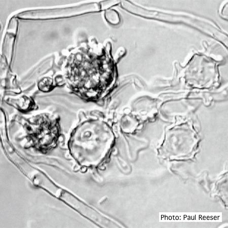 |
P. pseudotsugae sporangium 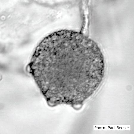 Broadly ovoid, papillate sporangium in water |
P. pseudotsugae colony morphology on V8 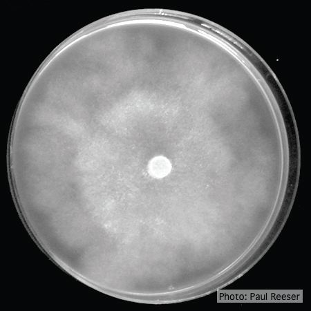 P. pseudotsugae colony growth on V8 agar |
|
P. pluvialis on Pinus radiata in New Zealand  Typical red needle cast symptoms along a twig. Lesions begin at the base of the needle which subsequently turns brown and is cast from the twig. |
P. pluvialis colony morphology on carrot agar  Colony morphology on carrot agar at 20 days |
P. megakarya oospore
P. megakarya oogonia, oospore, and antheridium
|
|
P. ramorum leaf symptoms on tan oak 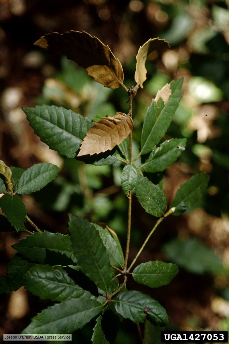 Tip symptoms on tanoak seedling (Notholithocarpus densiflorus). |
P. pinifolia on Pinus radiata 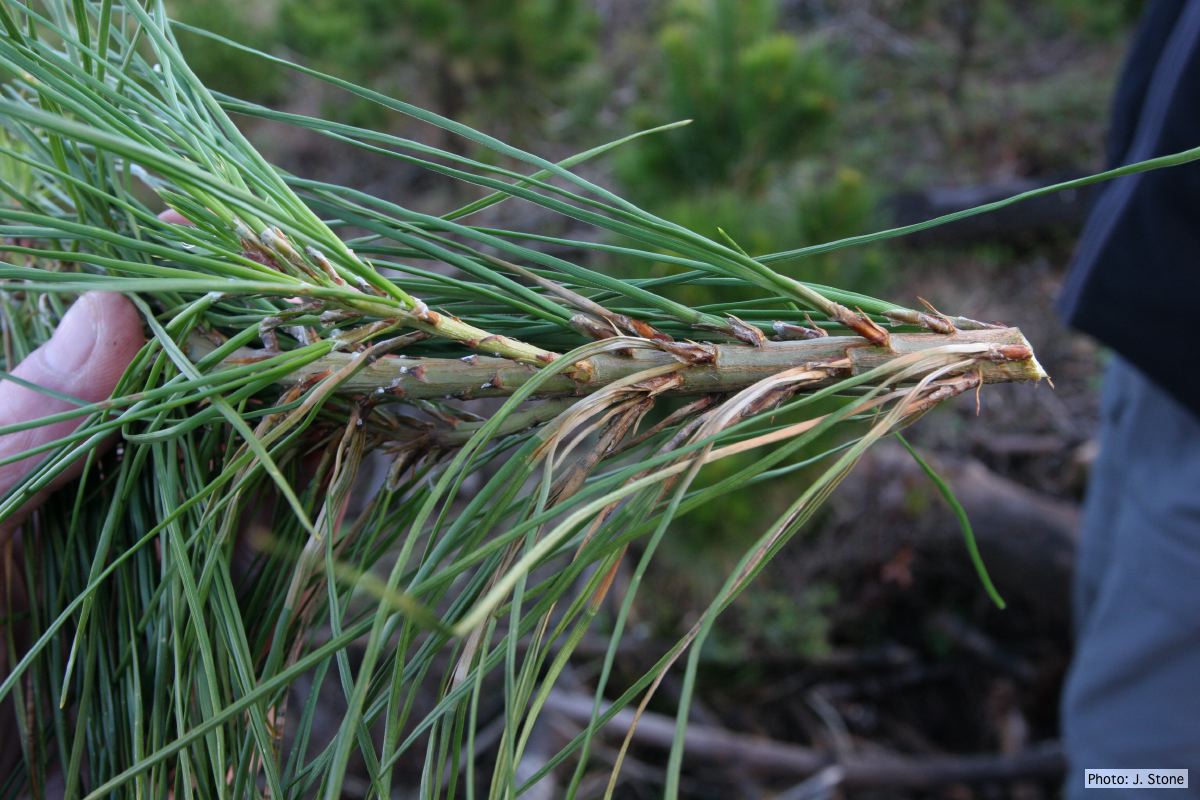 Pinus radiata, note grey and collapsed needle bases |
P. agathidicia disease symptoms on kauri 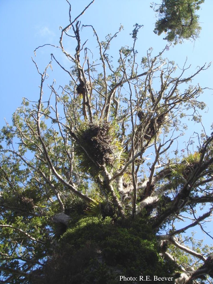 Crown decline of mature kauri, with branchlets with little or no leaves |
|
P. ramorum zoospores 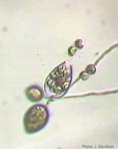 Sporangium of P. ramorum releasing zoospores |
P. megakarya sporangium 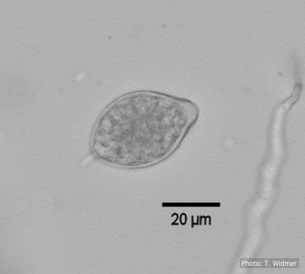 Caducous papillate sporangium of P. megakarya |
P. cambivora sporangium 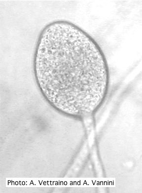 Ovoid non-papillate sporangia with well-rounded base and simple sporangiophore |