

P. siskiyouensis sporangium with lateral semi-papilla and subterminal, sub-basal insertion in the sporangiophore
Photo Gallery
Site will be retired 9/1/2026
This site is no longer being developed and will be retired on September 1, 2026. Please contact us if you have any questions or would like to provide support to continue the project.
|
P. siskiyouensis sporangium  |
Growth of P. palmivora on CMA 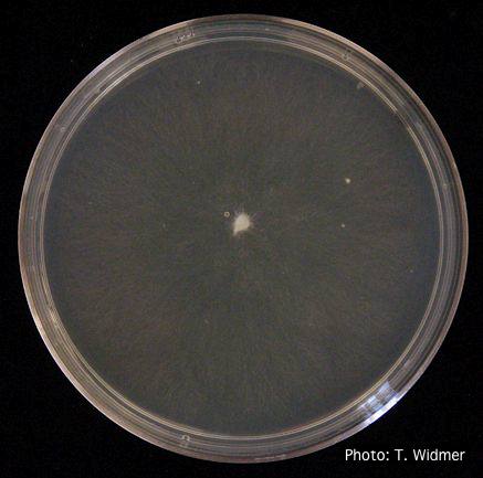 Growth of P. palmivora on corn meal agar |
P. agathidicida growth on MEA  Diffuse, non-patterned, colony morphology of ICMP 16471 (the original “Gadgil isolate”) after 10-days incubation at 20°C in the dark |
|
P. nemorosa hyphal swellings 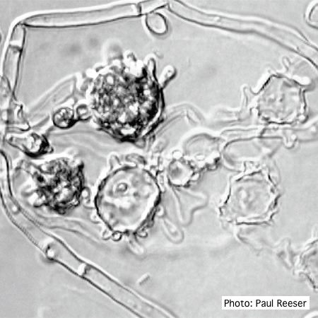 ‘Blistered’ hyphal swellings in agar |
P. cactorum sporangia 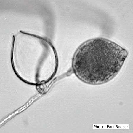 Broadly ovoid, papillate sporangia in water. |
P. pluvialis hyphal swellings 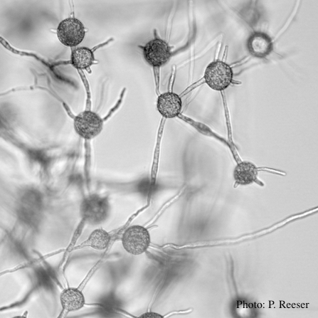 P. pluvialis hyphal swellings in water |
|
Black pod disease of cacao caused by P. palmivora 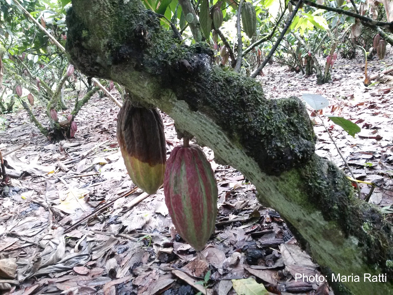 Black pod of cacao in Ecuador caused by P. palmivora (see lesioned fruit on left). |
P. siskiyouensis disease symptoms on Italian alder 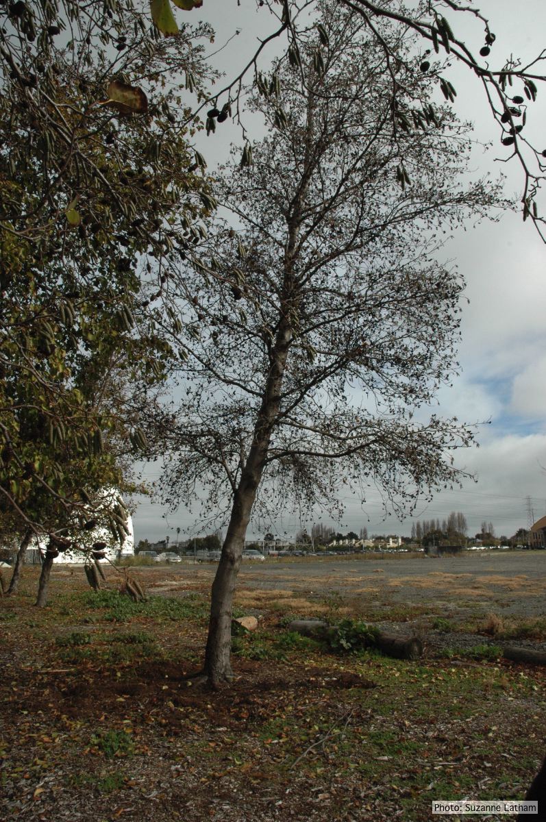 Phytophthora collar rot on Italian alder trees: standing, dead tree |
P. nemorosa oogonium 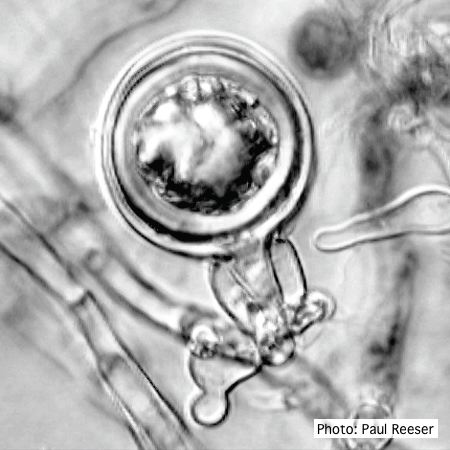 Oogonium with amphigynous antheridium |
|
P. cactorum colony morphology on PDA 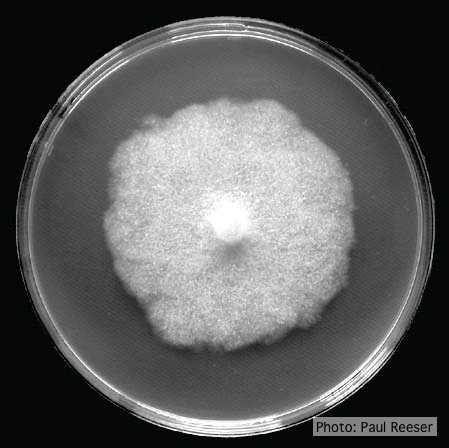 Colony morphology on PDA at 14 days |
P. cambivora colony morphology on PDA  Cottony colony morphology at 14 days at 20°C on PDA |
P. cactorum bleeding canker 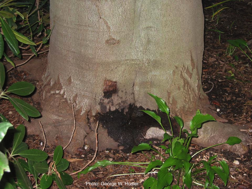 Bleeding canker on European beech (Fagus sylvatica) |