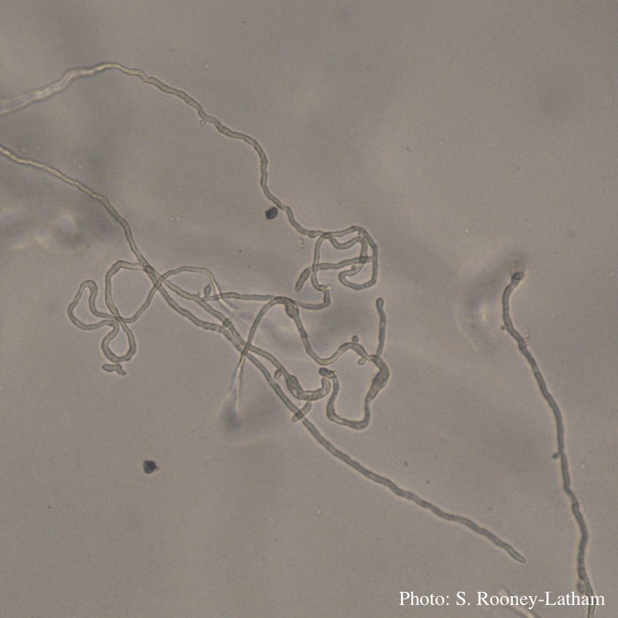
P. tentaculata hyphae
Looping hyphae commonly seen with P. tentaculata on PARP media
Photographer:
Suzanne Rooney-Latham
Pathogen Morphology:
Hyphal swellings
Scale:
Microscopic


Looping hyphae commonly seen with P. tentaculata on PARP media
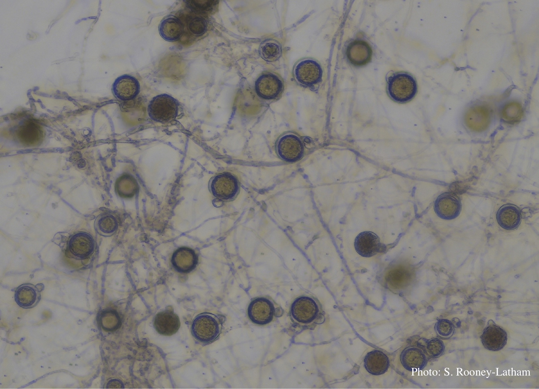
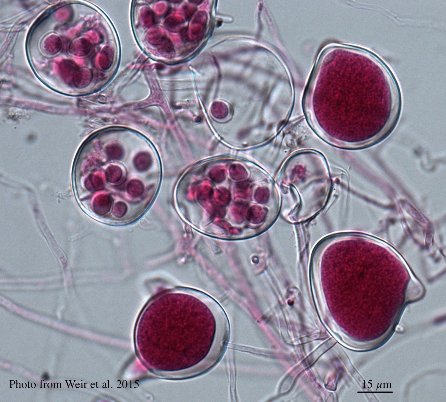
Oospores and oogonia with mostly paragynous but some amphigynous antheridia of P. tentaculata
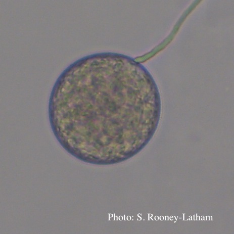
Terminal chlamydospore of P. tentaculata
Oospores in the roots of kauri seedlings inoculated with P. agathidicida. The root has been cleared with potassium hydroxide and bleached with peroxide before being stained with trypan blue (scale bar =100 µm).

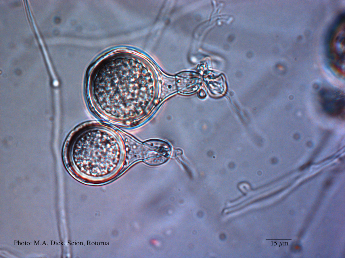
Globose to ovoid-ellipsoid, papillate sporangium
Oospores of P. agathidicida in the roots of kauri seedlings inoculated with P. agathidicida. The root has been cleared with potassium hydroxide and bleached with peroxide, before being stained with Trypan Blue

Comparative gametangial morphology of Phytophthora Clade 5 species, with SEM (top) and light microscopy (bottom). P. heveae has smooth walled oogonia with funnel-shaped, amphigynous antheridia. P. agathidicida has mildly stipulate oogonia with globose amphigynous antheridia. P.cocois has mildly bullate oogonia with reflexed amphigynous antheridia. P. castaneae has coarsely bullate oogonium with rugose protuberances and narrow amphigynous antheridia (Weir et al. 2015).

Differentiation of the cytoplasm within papillate sporangia into acid fuchsin stained zoospores
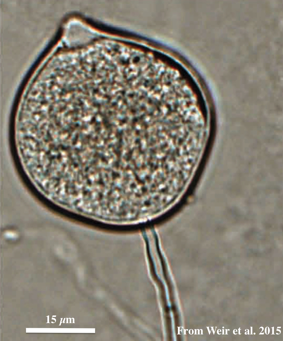
Light micrograph of P. agathidicida oospore (Scale bar equals 15 µm)
P. pluvialis sporangia on tape peel from infected Douglas-fir needle.
