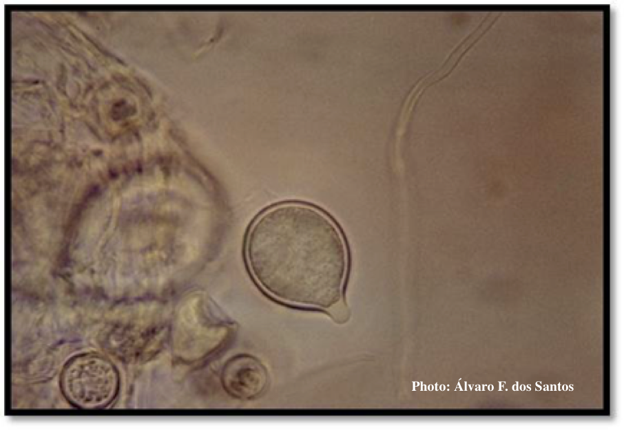
P. boehmeriae sporangia
Sporangia showing ovoid and ovoid to spherical shape and papillate condition

Photographer:
Álvaro F. dos Santos
Pathogen Morphology:
Sporangia
Scale:
Microscopic


Sporangia showing ovoid and ovoid to spherical shape and papillate condition
Sporangium showing ovoid and ovoid to spherical shape and papillate condition
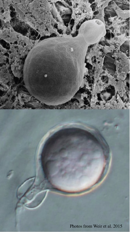
P. agathidicida oogonia with SEM (top) and light microscopy (bottom)
Clusters of sporangia emerge from stomata of an infected radiata pine needle.

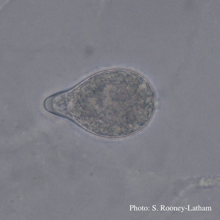
Papillate sporangium of P. tentaculata
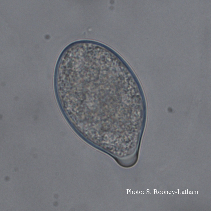
Papillate sporangium of P. tentaculata

P. tentaculata chlamydospore with short hyphal projection

Papillate sporangium of P. tentaculata with an elongated neck or beak.
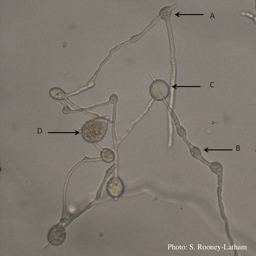
Hyphal swellings occuring at branching points of Mycelium (A), Intercalary hyphal swellings (B), Chlamydospore (C ), Sporangia (D)
Paragynous antheridium attached to oogonium with oospore
