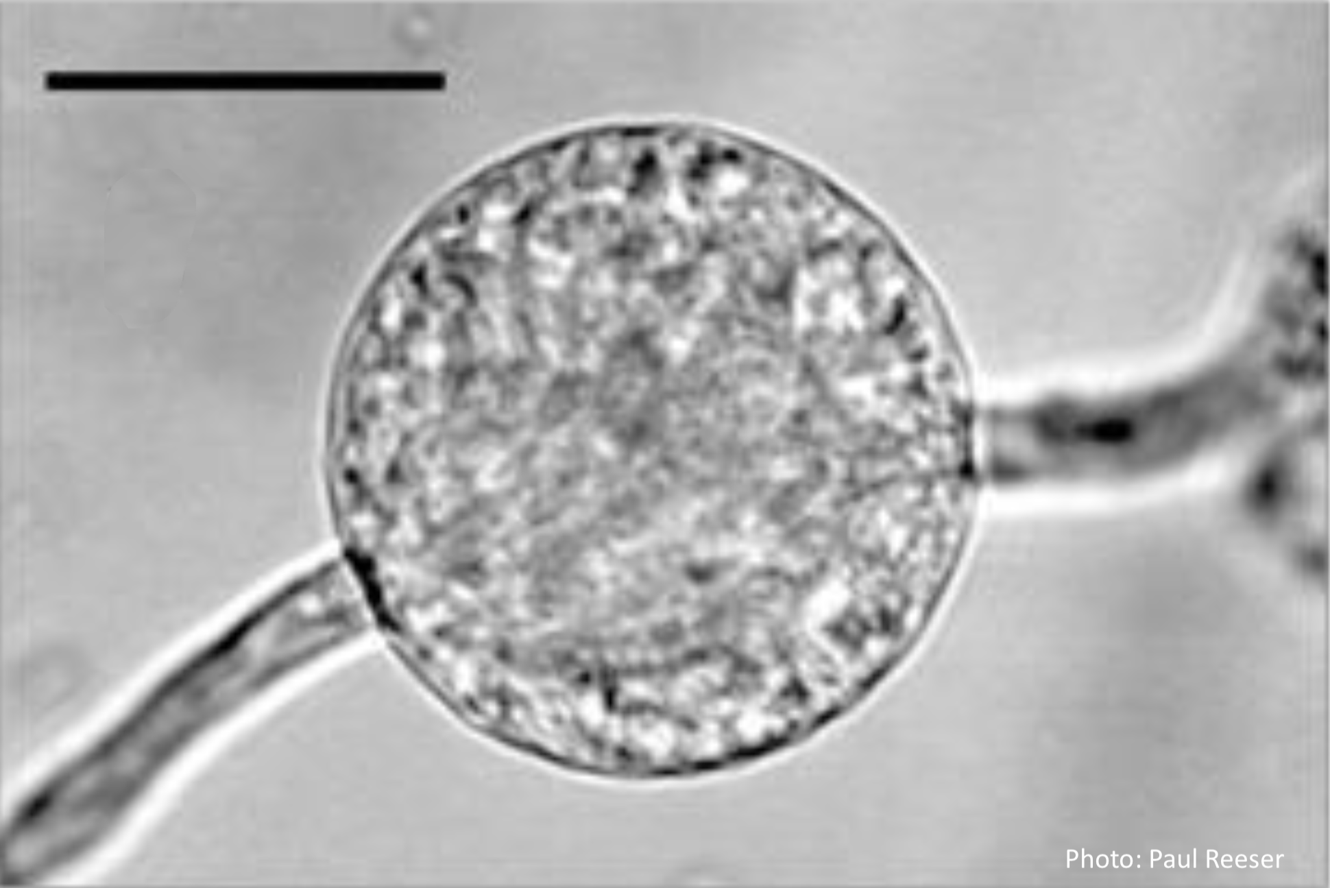
P. chlamydospora sporangium
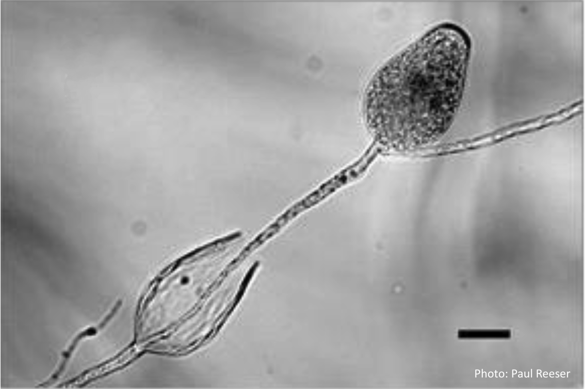
Phytophthora chlamydospora sporangia in water, showing internal proliferation. Bar is 20 µm.
Photographer:
Paul Reeser
Pathogen Morphology:
Sporangia
Scale:
Microscopic


Phytophthora chlamydospora sporangia in water, showing internal proliferation. Bar is 20 µm.
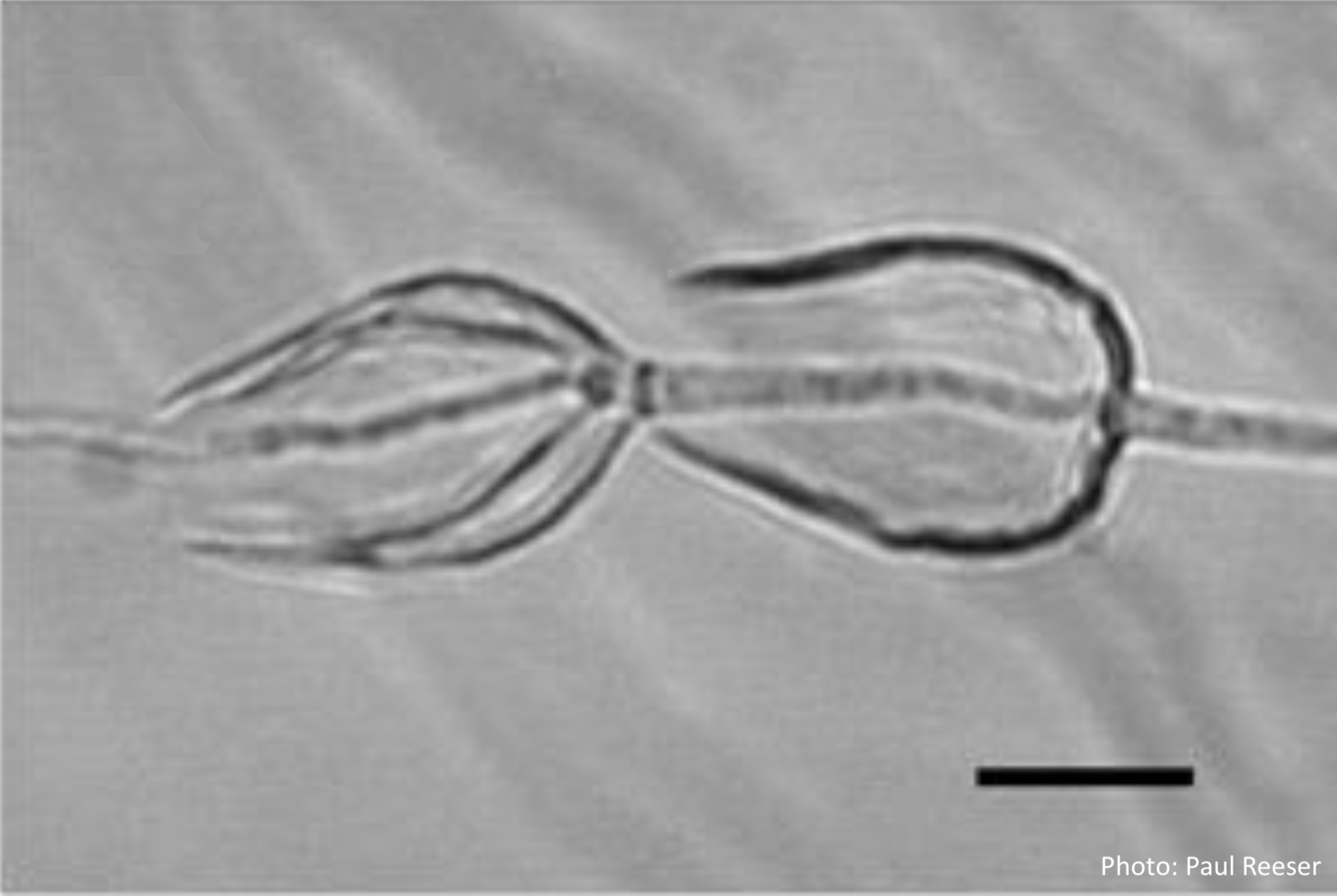
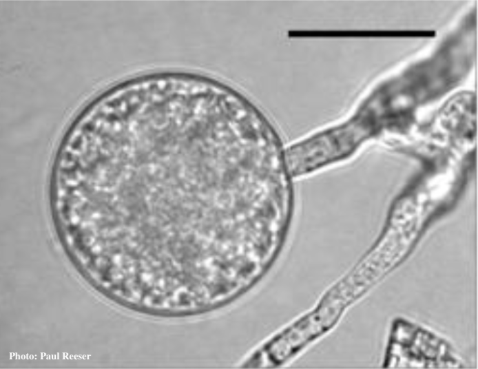
Phytophthora chlamydospora sporangia in water, showing subsporangial elongation. Bar is 20 µm.
Phytophthora chlamydospora sporangium in water. Bar is 20µm.
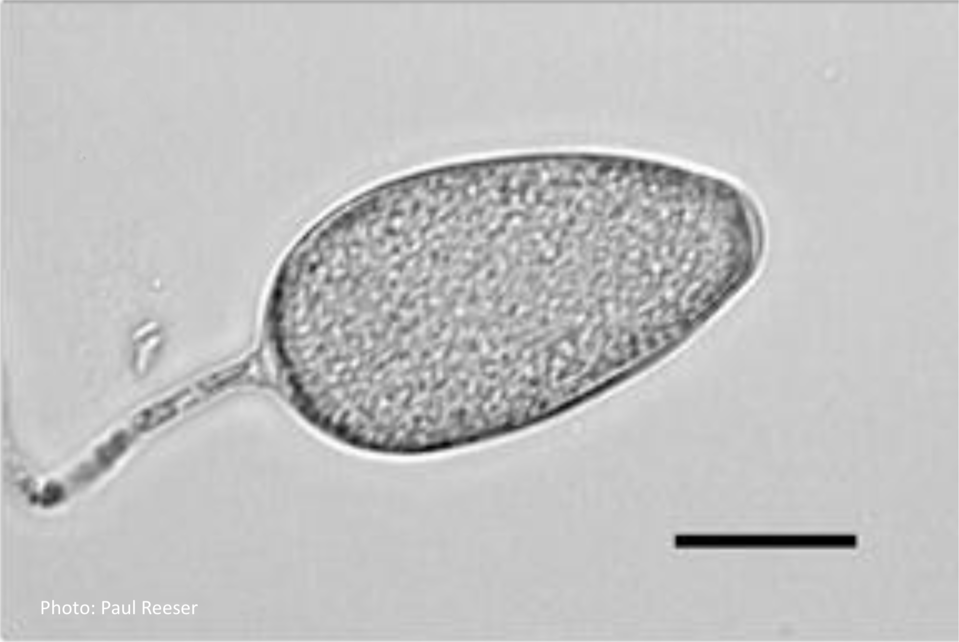
Phytophthora chlamydospora sporangium in water. Bar is 20µm.

Phytophthora chlamydospora chlamydospore in agar. Bar is 20µm.
Phytophthora chlamydospora chlamydospore in agar. Bar is 20µm.
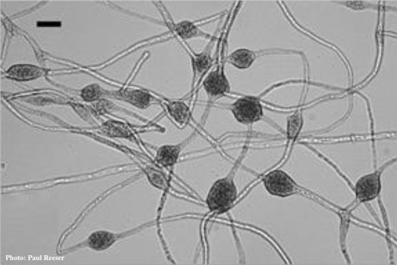
Phytophthora chlamydospora chlamydospore in agar. Bar is 20µm.

Phytophthora chlamydospora chlamydospore in agar. Bar is 20 µm.
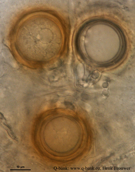
P. nicotianae overview of sporangia 40x. Photo from Q-bank: www.q-bank.eu, Henk Brouwer (CBS-KNAW, Utrecht, The Netherlands)

P. nicotianae oogonia 100x. Photo from Q-bank: www.q-bank.eu, Henk Brouwer (CBS-KNAW, Utrecht, The Netherlands)