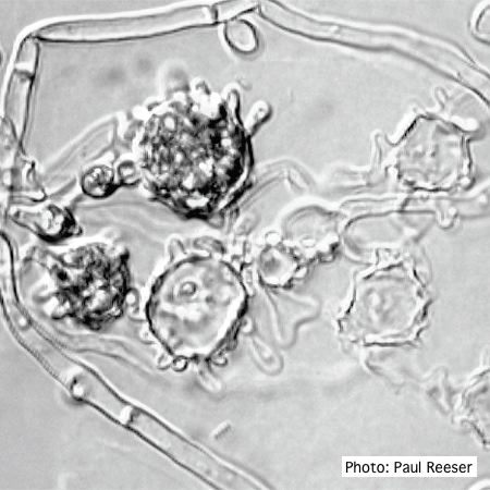
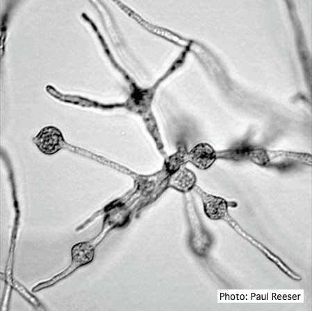
P. nemorosa hyphal swellings
‘Blistered’ hyphal swellings in agar
Photographer:
P. Reeser
Pathogen Morphology:
Hyphal swellings
Microscopic photos
Scale:
Microscopic


‘Blistered’ hyphal swellings in agar
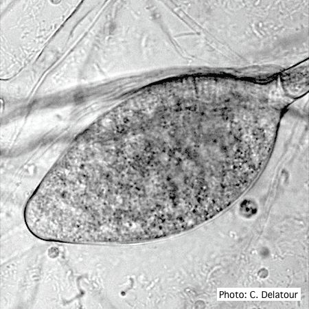
Ovoid, semi-papillate sporangium showing medium length pedicel.

Ovoid, semi-papillate sporangia showing sympodial development of sporangiophore

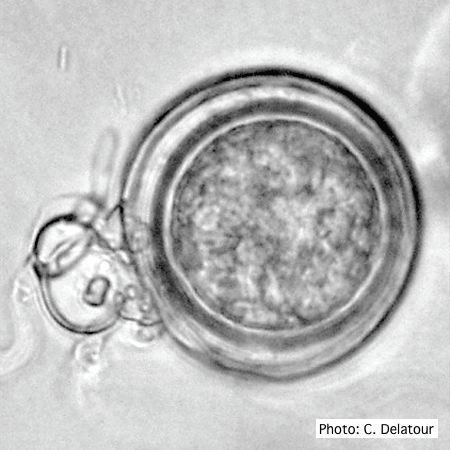
Oogonium with paragynous antheridia applied close to the ogonial stalk.
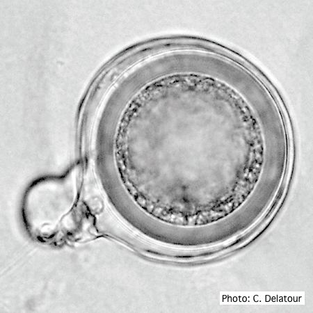
Oogonium with paragynous antheridia applied close to the ogonial stalk.
Ovoid, non-papillate sporangia

Ovoid, non-papillate sporangia showing internal proliferation of sporangiophore
Cluster of small, angular to globose hyphal swellings formed in water
Sporangiophore showing internal proliferation through empty sporangia after zoospore release

Ovoid non-papillate sporangia in water.
