

Pinus radiata needles showing colour changes following infection with red needle cast disease. The tissues around the initial infection at the base or along the needle senesce, and change yellow and then brown as indicated by the arrows before the needles cast.
Photo Gallery
Site will be retired 9/1/2026
This site is no longer being developed and will be retired on September 1, 2026. Please contact us if you have any questions or would like to provide support to continue the project.
|
P. pluvialis on Pinus radiata in New Zealand  |
P. katsurae growth morphology on β-CMA 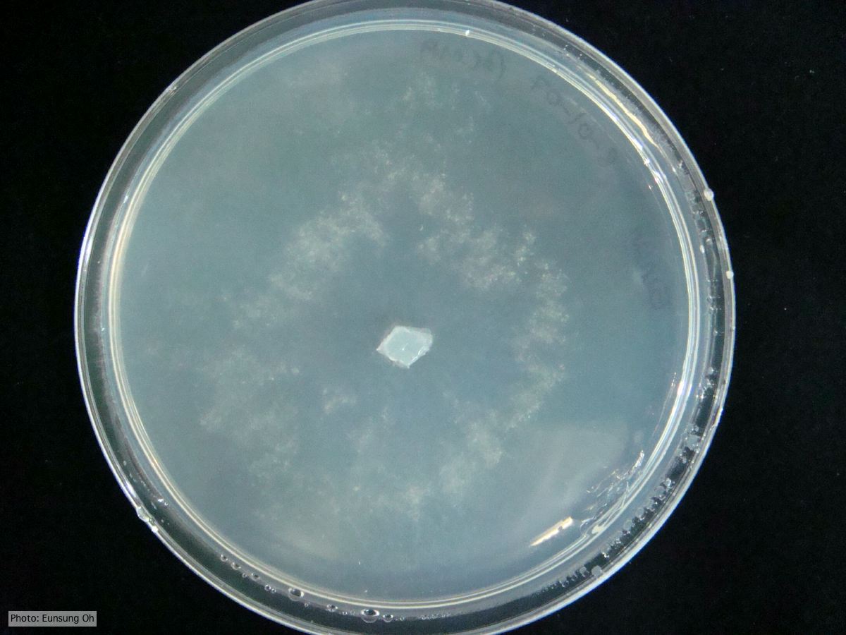 Growth morphology at 7 days on β-CMA |
P. chlamydospora sporangium 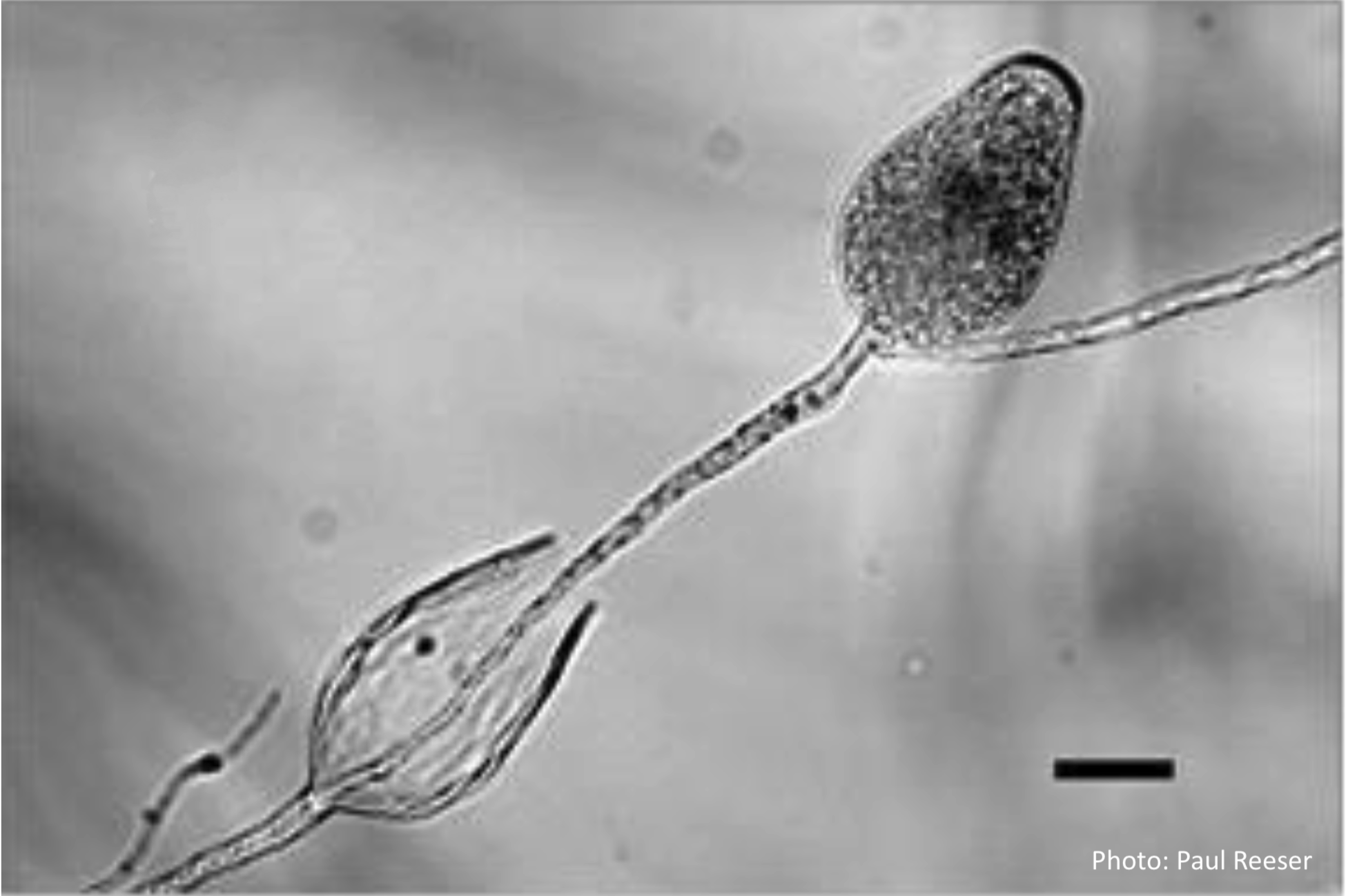 Phytophthora chlamydospora sporangia in water, showing subsporangial elongation. Bar is 20 µm. |
|
P. tentaculata oogonia and antheridia 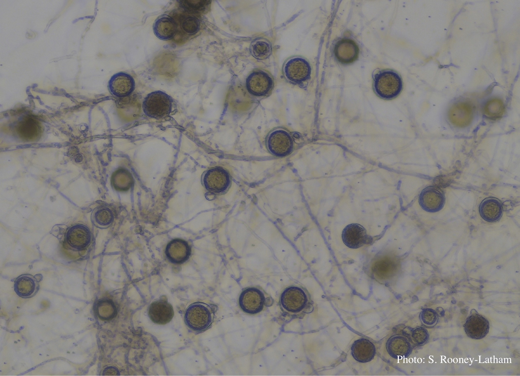 Oospores and oogonia with mostly paragynous but some amphigynous antheridia of P. tentaculata |
P. lateralis on Port Orford cedar  Localized branch infection of Chaemacyparis lawsoniana in Lopérec, France |
P. alni symptoms on European Alder 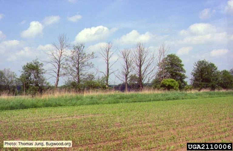 Mature, riparian common alder (A. glutinosa) stand with high impact of Phytophthora root and collar rot. |
|
P. cambivora colony morphology on PDA 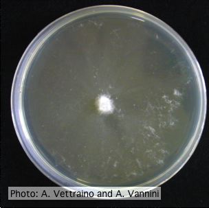 Appressed colony morphology at 14 days at 20°C on PDA |
P. cinnamomi on Banksia 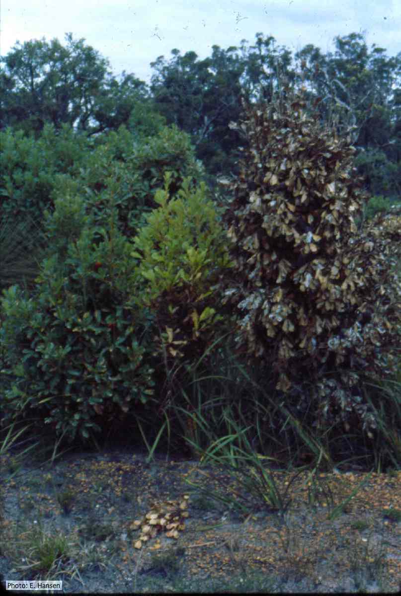 Dead and dying Banksia, Western Australia |
P. cambivora disease symptoms 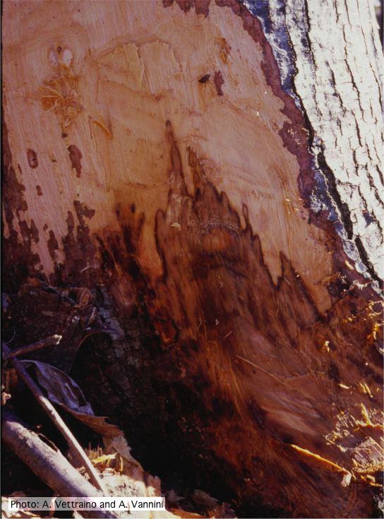 Collar canker rot of Ink disease on sweet chestnut |
|
P. cambivora sporangium with nested proliferation 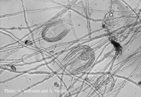 Empty sporagia showing internal nested proliferation |
P. cambivora colony morphology on MA  Appressed colony morphology at 14 days at 20°C on MA |
P. cinnamomi cork oak decline  Red oak with embedded lesions, France |