

Ovoid non- papillate sporangia
Photo Gallery
Site will be retired 9/1/2026
This site is no longer being developed and will be retired on September 1, 2026. Please contact us if you have any questions or would like to provide support to continue the project.
|
P. cambivora sporangium 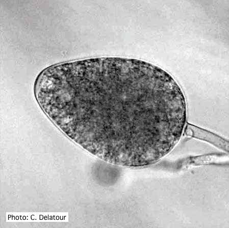 |
P. ramorum zoospores 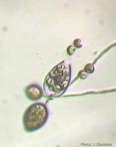 Sporangium of P. ramorum releasing zoospores |
P. pinifolia sporulation 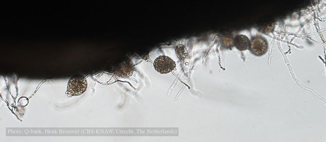 Sporulation on edge of hemp seed, photo from Q-bank, used with permission. |
|
P. agathidicida sporangium 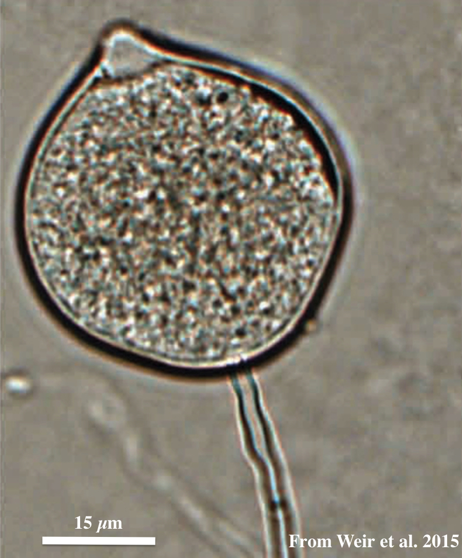 Globose to ovoid-ellipsoid, papillate sporangium |
P. pinifolia hyphal growth 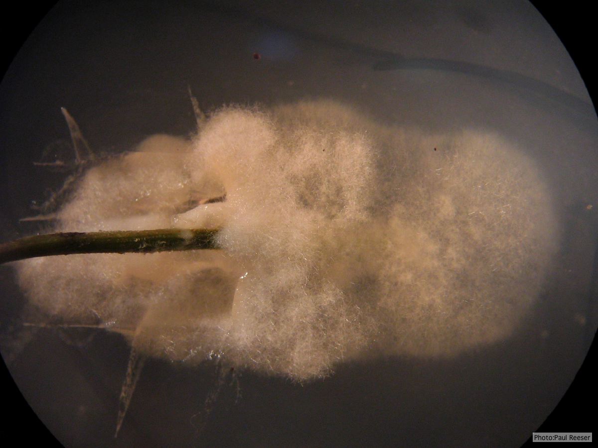 P. pinifolia pathogen growing from infected needle on selective agar |
P. cambivora tar spots 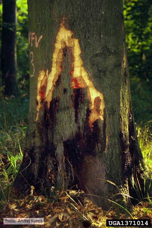 Tar spots on European beech (Fagus sylvatica) with bark removed. Lesse, Germany |
|
P. cactorum bleeding canker 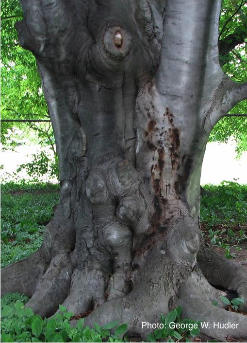 Bleeding canker on European beech (Fagus sylvatica) |
P. cambivora colony morphology on PDA 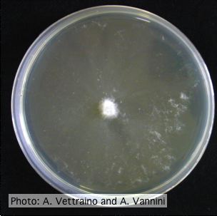 Appressed colony morphology at 14 days at 20°C on PDA |
P. agathidicia sporangia 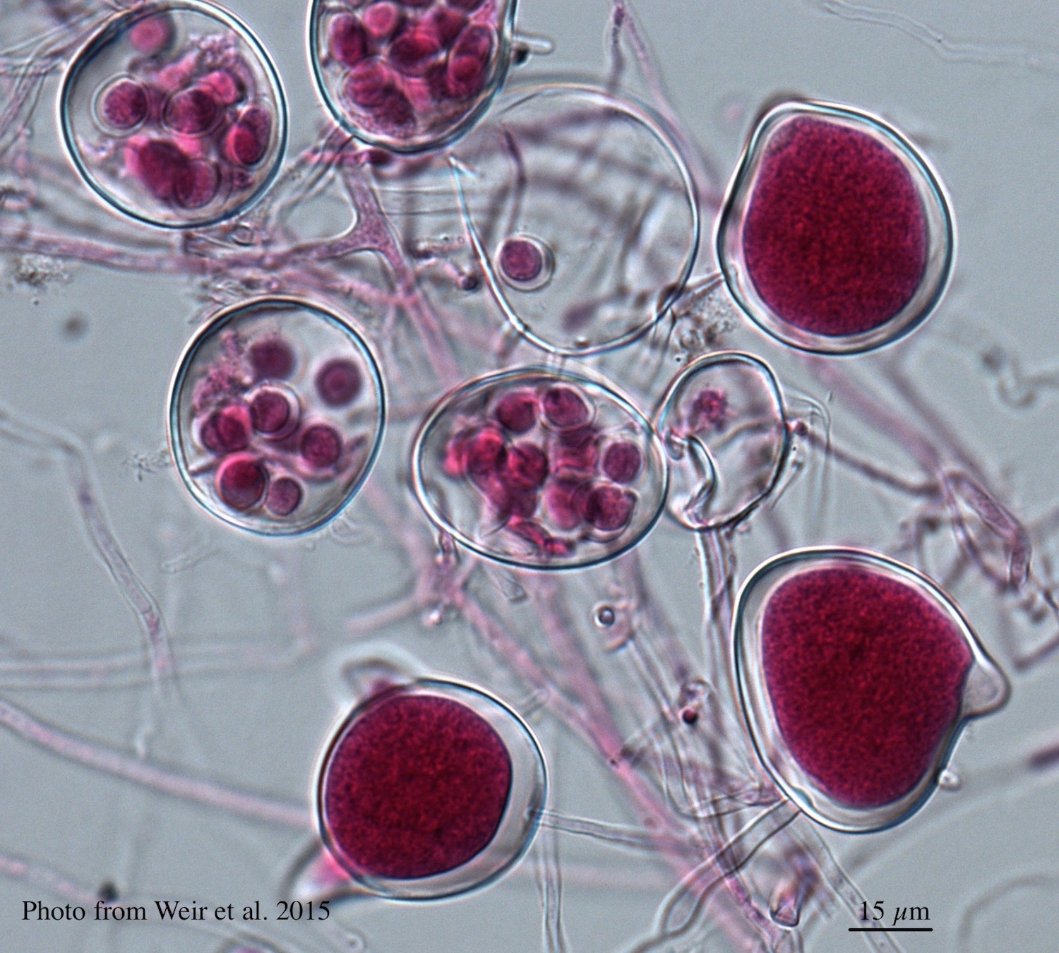 Differentiation of the cytoplasm within papillate sporangia into acid fuchsin stained zoospores |
|
P. pluvialis on Pinus radiata needle  Clusters of sporangia emerge from stomata of an infected radiata pine needle. |
P. cryptogea colony morpholgy on PDA 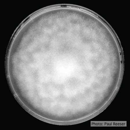 Colony morphology on PDA at 14 days |
P. austrocedrae colony morphology on Tomato juice agar 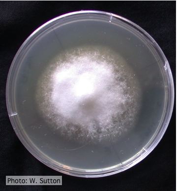 Colony morphology of P. austrocedrae at 16 ºC after 4 weeks on Tomato juice agar |