

Colony morphology on PDA at 14 days
Photo Gallery
Site will be retired 9/1/2026
This site is no longer being developed and will be retired on September 1, 2026. Please contact us if you have any questions or would like to provide support to continue the project.
|
P. siskiyouensis colony morphology on PDA 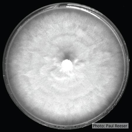 |
P. kernoviae oogonia 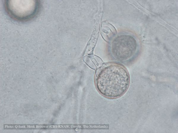 Oogonium with amphigynous antheridia, photo from Q-bank, used with permission |
P. pluvialis symptoms on Douglas-fir  Red needle cast symptoms on Douglas-fir in western Oregon, 2015 |
|
P. pseudosyringae colony morphology on PDA  Colony morphology on PDA at 14 days |
P. siskiyouensis canker on Italian alder 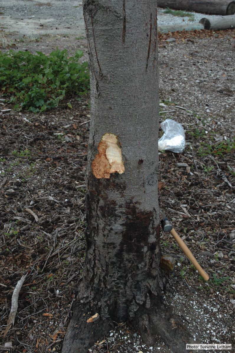 Bole lesions in the tissues under the bark of a bleeding canker: distinct margin between healthy and disease tissues |
P. austrocedrae hyphal swellings 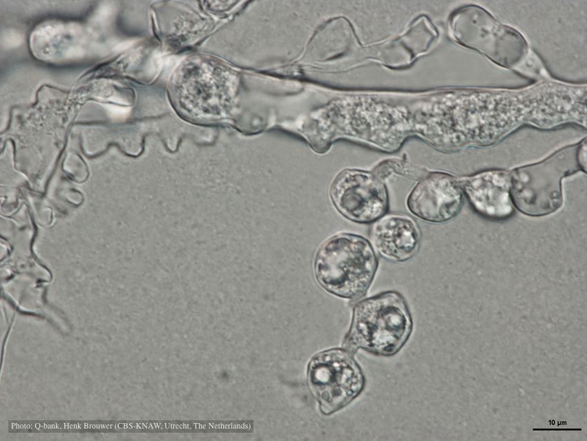 Hyphal swelling photo used with permission from Q-bank |
|
P. cambivora colony morphology on PDA 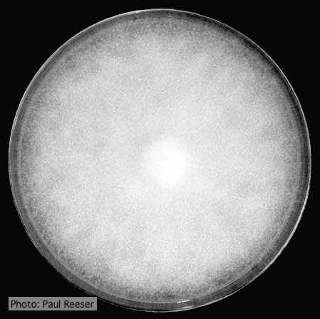 Colony morphology on PDA at 14 days |
P. ramorum chlamydospores 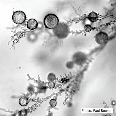 P. ramorum chlamydospores |
P. tentaculata sporangium 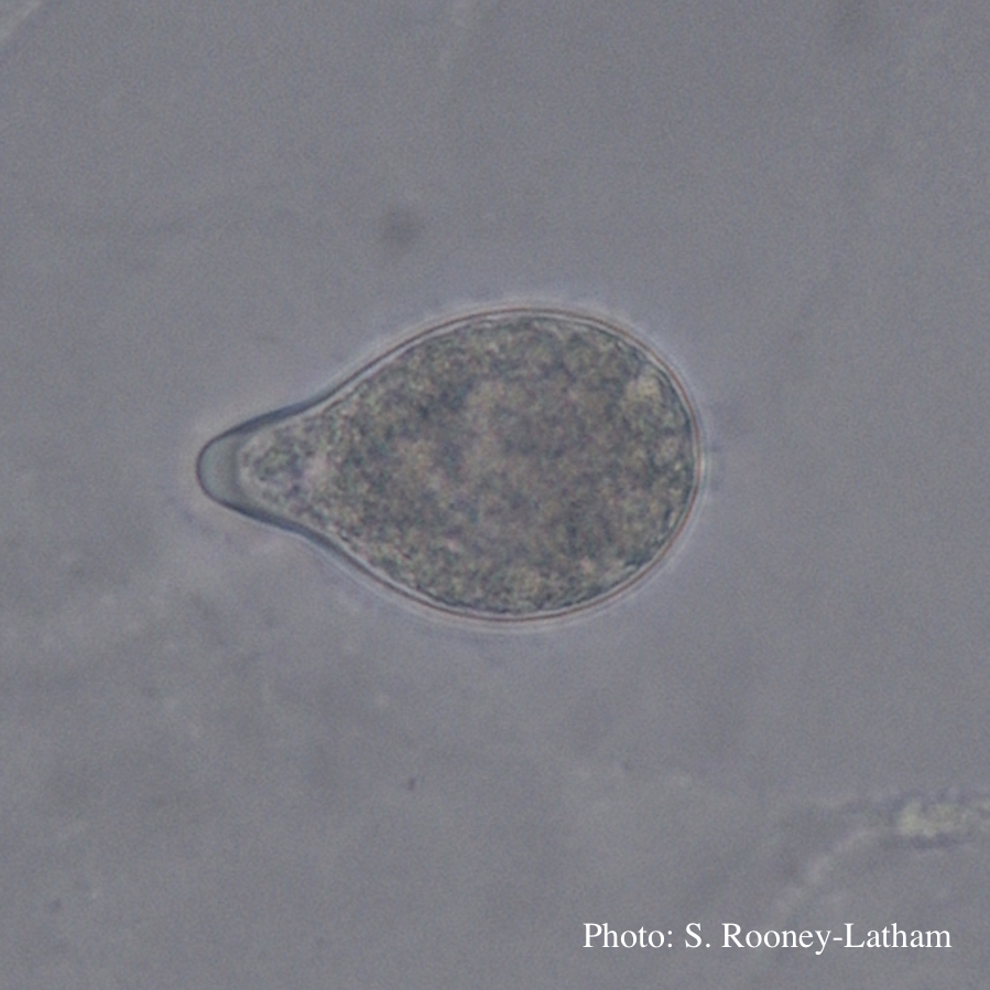 Papillate sporangium of P. tentaculata with an elongated neck or beak. |
|
P. tentaculata disease symptoms on sticky monkey flower 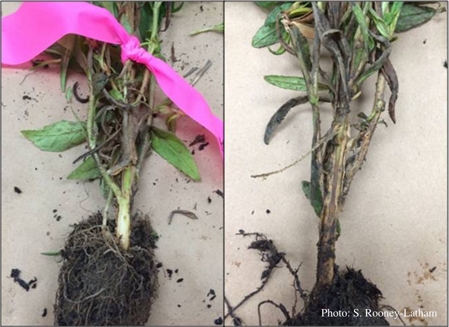 Crown and root rot (left) on sticky monkey flower (Diplacus aurantiacus) compared with a control (right) |
Port Orford cedar seedlings 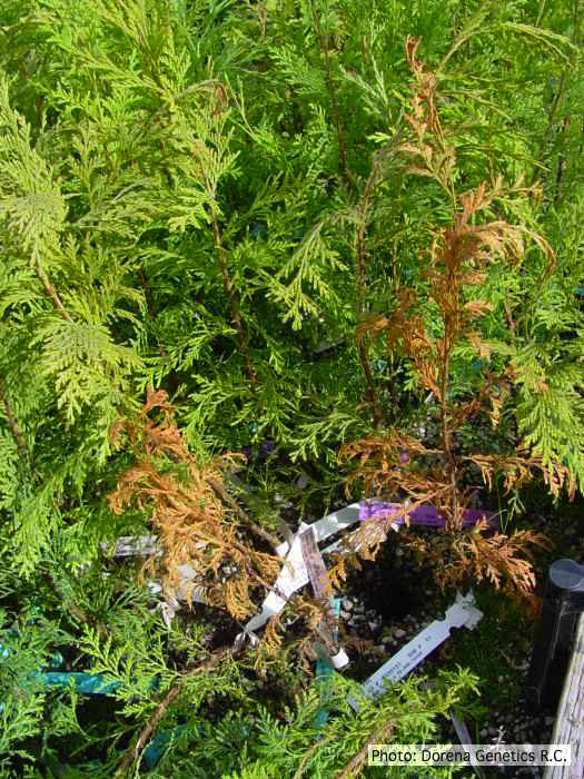 Raised beds for testing disease resistance of Port-Orford-cedar seedlings at the Dorena Genetic Resource Center |
P. cinnamomi on Banksia  Death of woodland Banksia, Western Australia |