

Sporangium with internal proliferation, photo from Q-bank, used with permission.
Photo Gallery
Site will be retired 9/1/2026
This site is no longer being developed and will be retired on September 1, 2026. Please contact us if you have any questions or would like to provide support to continue the project.
|
P. pinifolia sporangia 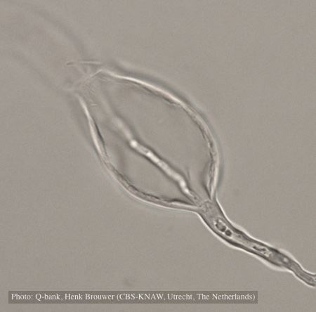 |
P. cambivora disease symptoms 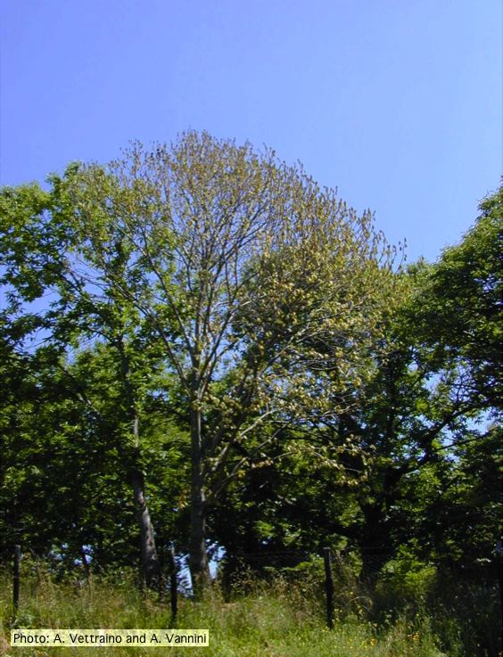 Crown symptoms of Ink disease on sweet chestnut |
P. cinnamomi hyphal swellings 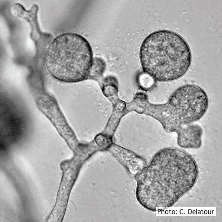 P. cinnamomi hyphal swellings (or thin walled chlamydospores) |
|
Chlamydospore of P. lateralis 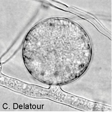 Terminal chlamydospore on a short side stalk |
P. austrocedrae colony morphology on Tomato juice agar with B sitosterol 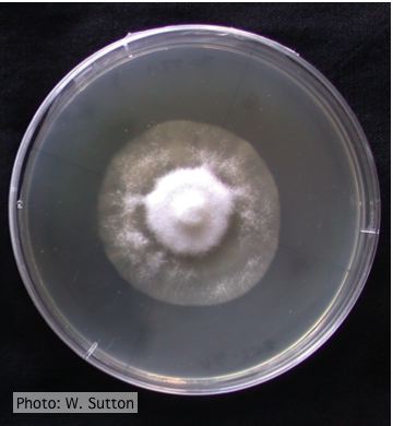 Colony morphology of P. austrocedrae at 16 ºC after 4 weeks on Tomato juice agar with B sitosterol |
P. cactorum bleeding canker 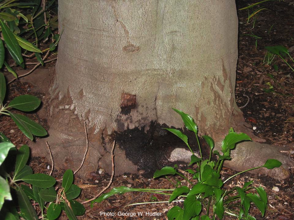 Bleeding canker on European beech (Fagus sylvatica) |
|
Growth of P. arenaria on half-strength PDA 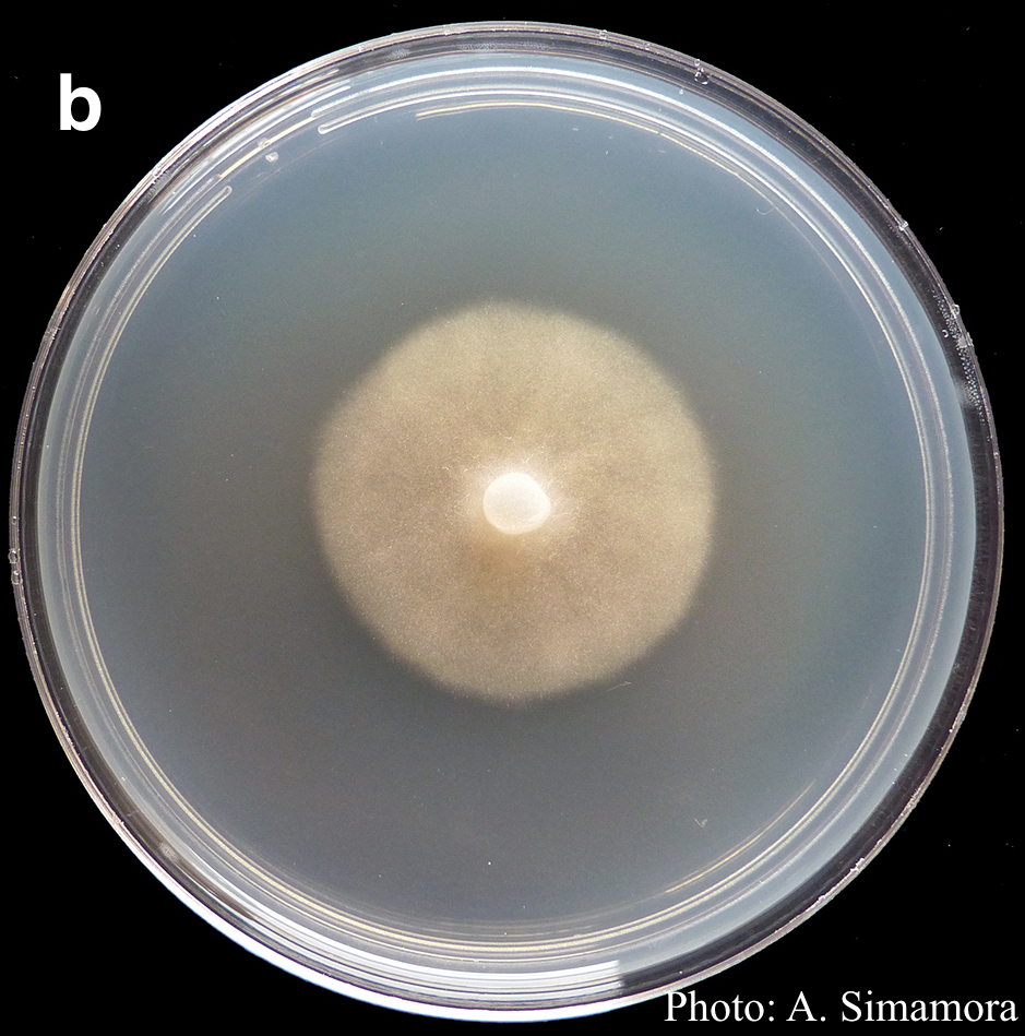 Colony morphology of Phytophthora arenaria after 7 days at 20°C on half-strength PDA |
P. tentaculata disease symptoms on California mugwort  Nursery grown California mugwort plant (Artemisia douglasiana) infected with P. tentaculata and exhibiting severe root and crown rot |
P. cinnamomi on Jarrah 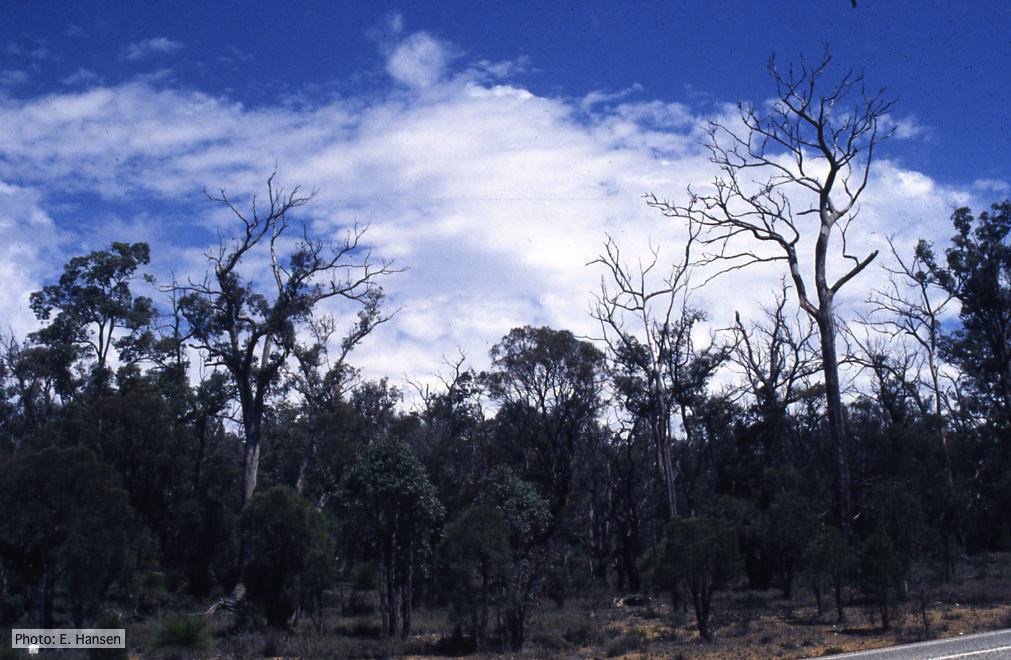 Dieback in Jarrah, Western Australia |
|
Port Orford Cedar Hedge row  Chamaecyparis lawsoniana residential hedge row with alive and dead trees |
P. kernoviae canker  Bole lesion on Fagus sylvatica |
P. agathidicida lesion on kauri tree 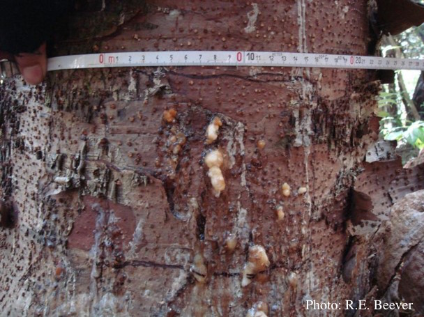 Close up of gum oozing out of lower trunk lesions of a young kauri tree at Maungaroa Ridge, Piha region of Waitakere Regional Park |