

P. siskiyouensis sporangium with lateral semi-papilla and subterminal, sub-basal insertion in the sporangiophore
Photo Gallery
Site will be retired 9/1/2026
This site is no longer being developed and will be retired on September 1, 2026. Please contact us if you have any questions or would like to provide support to continue the project.
|
P. siskiyouensis sporangium  |
P. cambivora sporangium with internal extended proliferation 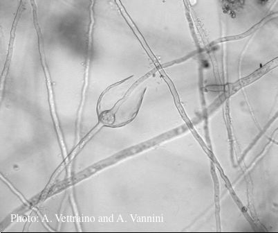 Empty sporangia showing internal extended proliferation |
P. pseudosyringae hyphal swellings  Sub-globose hyphal swellings in water |
|
P. cactorum bleeding canker 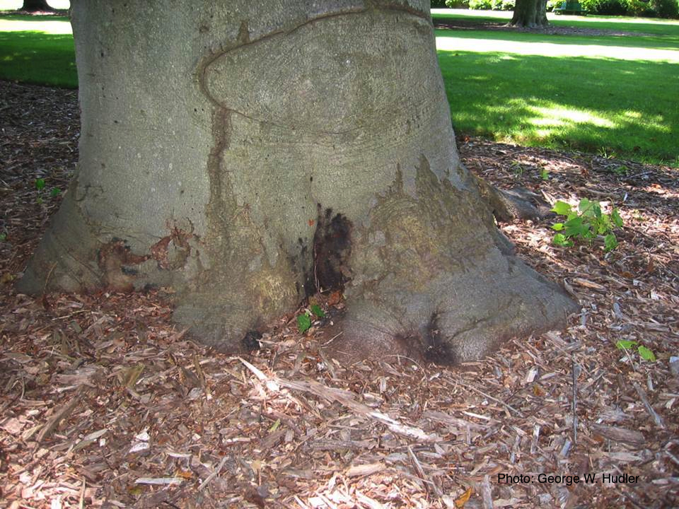 Bleeding canker on European beech (Fagus sylvatica) |
P. frigida sporangia  Noncaducous sporangia showing ovoid shape and papillate condition |
P. cambivora oogonium 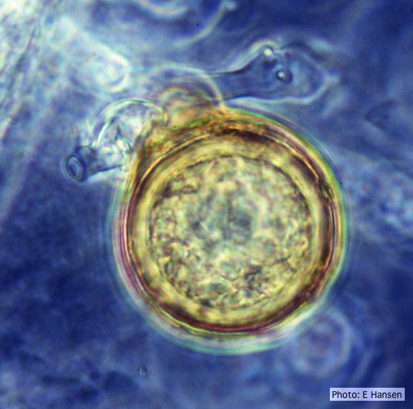 P. cambivora oogonium with antheridium |
|
P. pseudosyringae sporangium 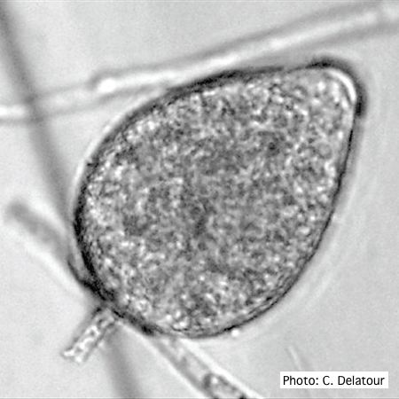 Ovoid, semipapillate sporangia showing medium length pedicel |
P. pluvialis - appearance of new growth 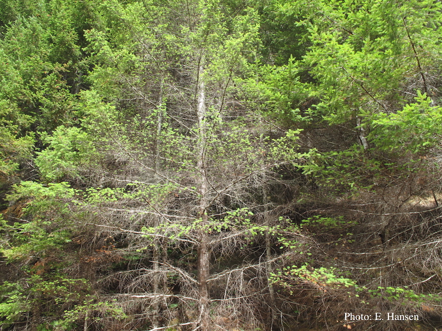 Tufted appearance of new growth from surviving buds on Douglas-fir, one year after defoliation. |
Port Orford cedar seedlings 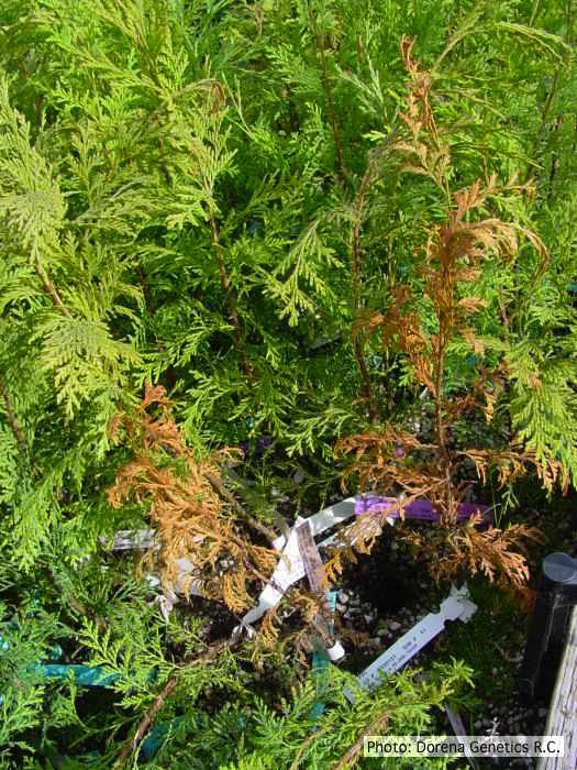 Raised beds for testing disease resistance of Port-Orford-cedar seedlings at the Dorena Genetic Resource Center |
|
P. austrocedrae colony morphology on Tomato juice agar 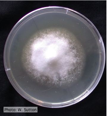 Colony morphology of P. austrocedrae at 16 ºC after 4 weeks on Tomato juice agar |
P. frigida chlamydospore 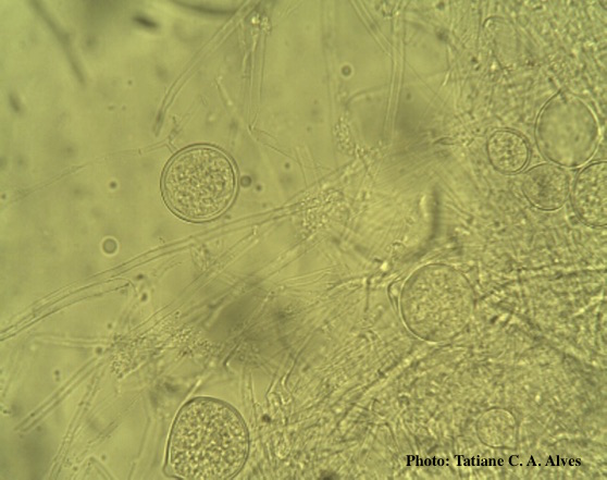 Globose chlamydospores of P. frigida |
P. pseudosyringae sporangia  Ovoid, semipapillate sporangia showing sympodial development of sporangiophore |