

Red needle cast symptoms on Douglas-fir in western Oregon, 2015
Photo Gallery
Site will be retired 9/1/2026
This site is no longer being developed and will be retired on September 1, 2026. Please contact us if you have any questions or would like to provide support to continue the project.
|
P. pluvialis symptoms on Douglas-fir 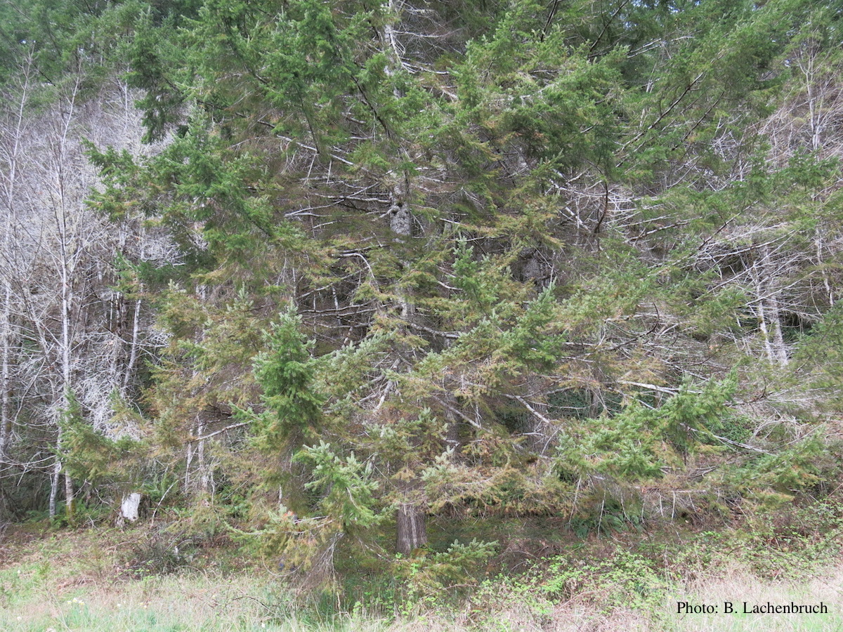 |
P. cinnamomi cork oak decline  Cork oak decline, Portugal |
P. siskiyouensis bleeding canker  Bole lesions in the tissues under the bark of a bleeding canker: discoloration in the secondary phloem tissue |
|
P. ramorum leaf symptoms on tan oak 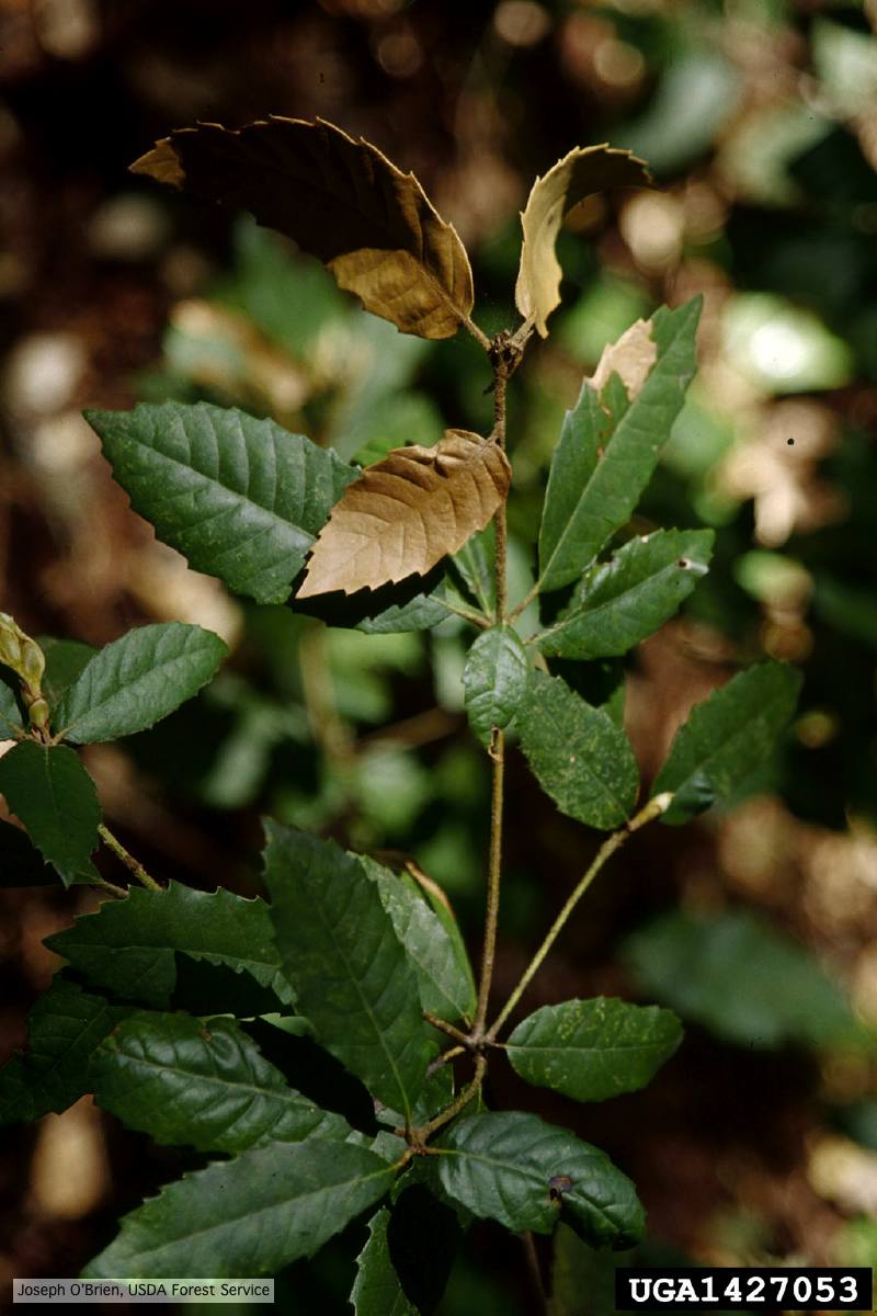 Tip symptoms on tanoak seedling (Notholithocarpus densiflorus). |
P. cinnamomi hyphal swellings 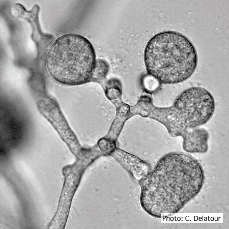 P. cinnamomi hyphal swellings (or thin walled chlamydospores) |
P. kernoviae leaf wilt 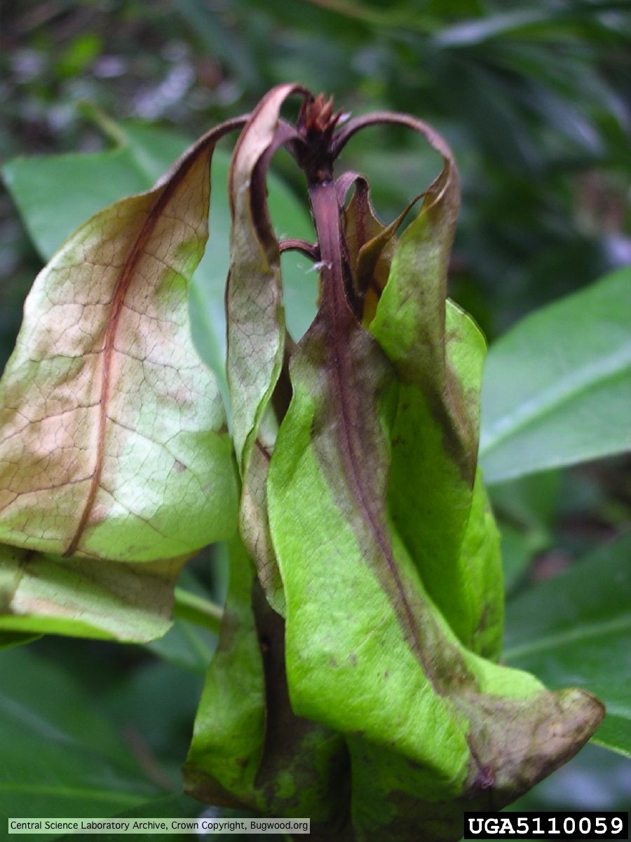 Necrosis of rhododendron leaves. |
|
P. katsurae disease symptoms  Infected chestnut (Castanea) with bleeding canker |
P. nemorosa sporangia  Ovoid, semi-papillate sporangia showing sympodial development of sporangiophore |
P. pluvialis - appearance of new growth 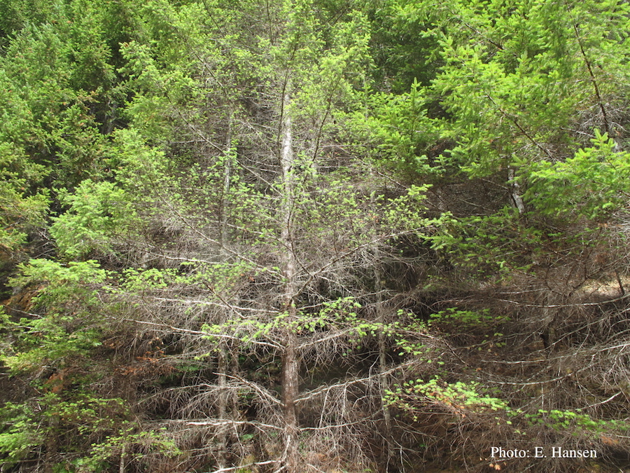 Tufted appearance of new growth from surviving buds on Douglas-fir, one year after defoliation. |
|
P. cinnamomi on Fraser fir 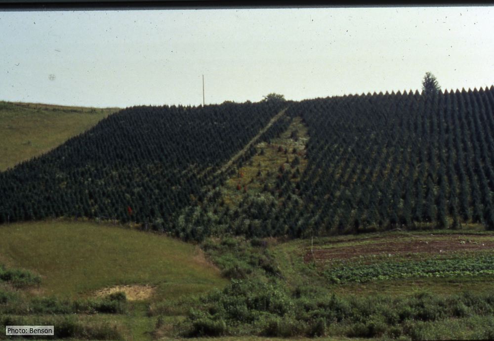 Frasier fir Christmas trees, North Carolina |
P. tentaculata sporangium  Papillate sporangium of P. tentaculata |
P. cambivora disease symptoms 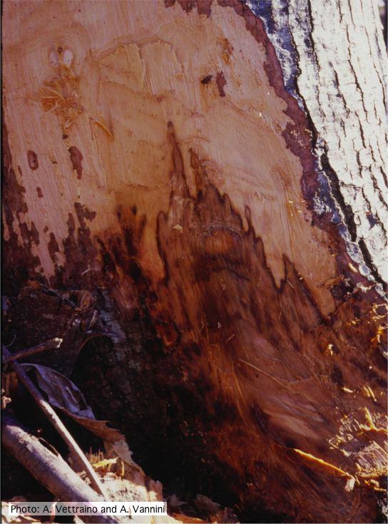 Collar canker rot of Ink disease on sweet chestnut |