

Colony morphology on PDA at 14 days
Photo Gallery
Site will be retired 9/1/2026
This site is no longer being developed and will be retired on September 1, 2026. Please contact us if you have any questions or would like to provide support to continue the project.
|
P. cambivora colony morphology on PDA 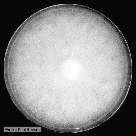 |
P. cinnamomi cork oak decline  Cork oak decline, Portugal |
P. tentaculata oogonia and antheridia 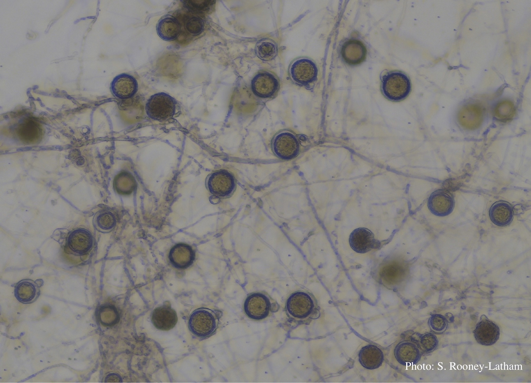 Oospores and oogonia with mostly paragynous but some amphigynous antheridia of P. tentaculata |
|
P. austrocedrae - hyphal swellings  Morphology of hyphae of Phytophthora austrocedrae, from Greslebin et al. 2007 |
P. cactorum bleeding canker 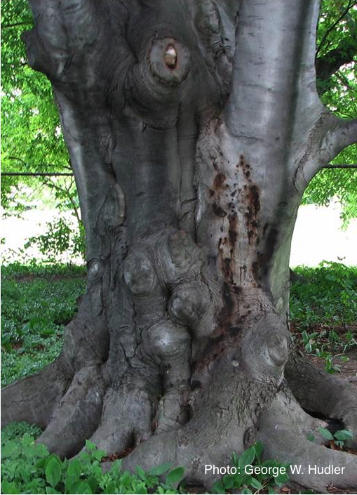 Bleeding canker on European beech (Fagus sylvatica) |
P. tentaculata disease symptoms on sticky monkey flower 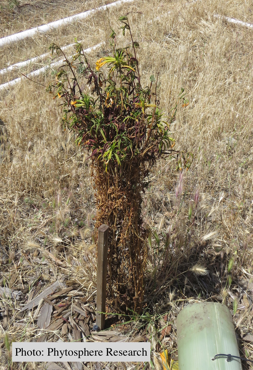 Outplanted sticky monkey flower (Diplacus aurantiacus) infected with P. |
|
P. lateralis sporangia  Sympodial sporangiophore with external proliferation |
P. cactorum colony morphology on V8  Colony morphology on V8 at 14 days |
P. kernoviae oogonia 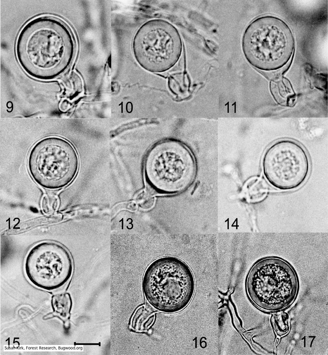 Mycol.Res 109, 853-859; Representative oogonia, antheridia and thick walled plerotic oospores of Phytophthora kernoviae. |
|
P. palmivora colony morphology on PDA 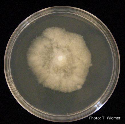 P. palmivora colony morphology on PDA |
P. pinifolia coenocytic hyphae 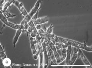 Coenocytic hyphae (from Duran et al. 2008). Scale bar = 20 μm. |
Phytophthora chlamydospora sporangium 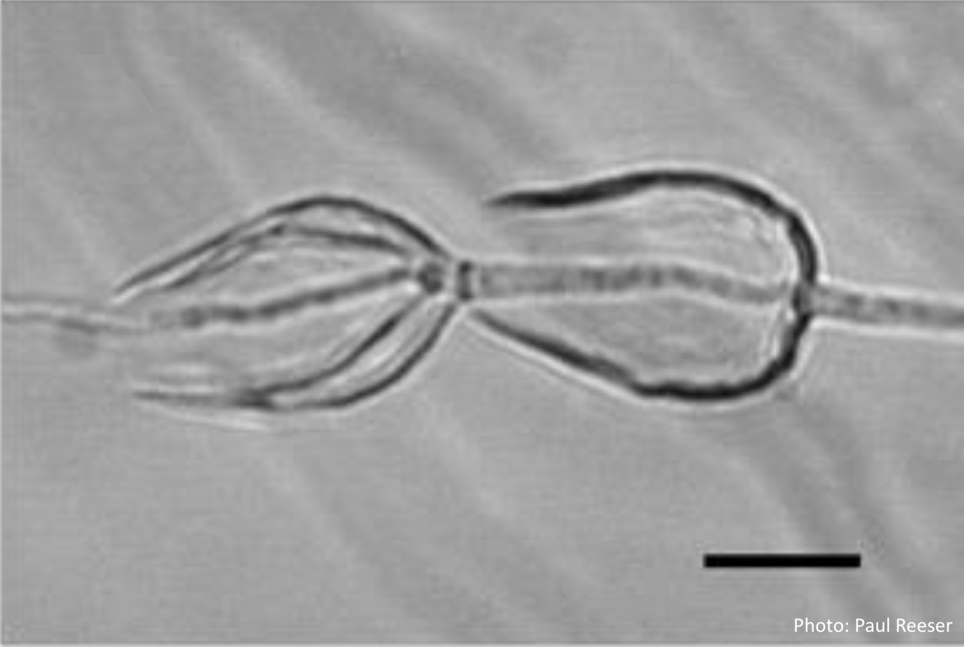 Phytophthora chlamydospora sporangia in water, showing internal proliferation. Bar is 20 µm. |