

Ovoid, semipapillate sporangia showing sympodial development of sporangiophore
Photo Gallery
Site will be retired 9/1/2026
This site is no longer being developed and will be retired on September 1, 2026. Please contact us if you have any questions or would like to provide support to continue the project.
|
P. pseudosyringae sporangia  |
P. cambivora disease symptoms 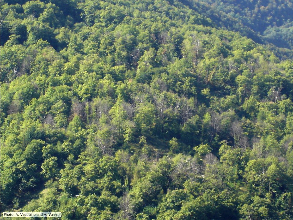 Ink disease impact in sweet chestnut forest in Italy |
P. tentaculata sporangium 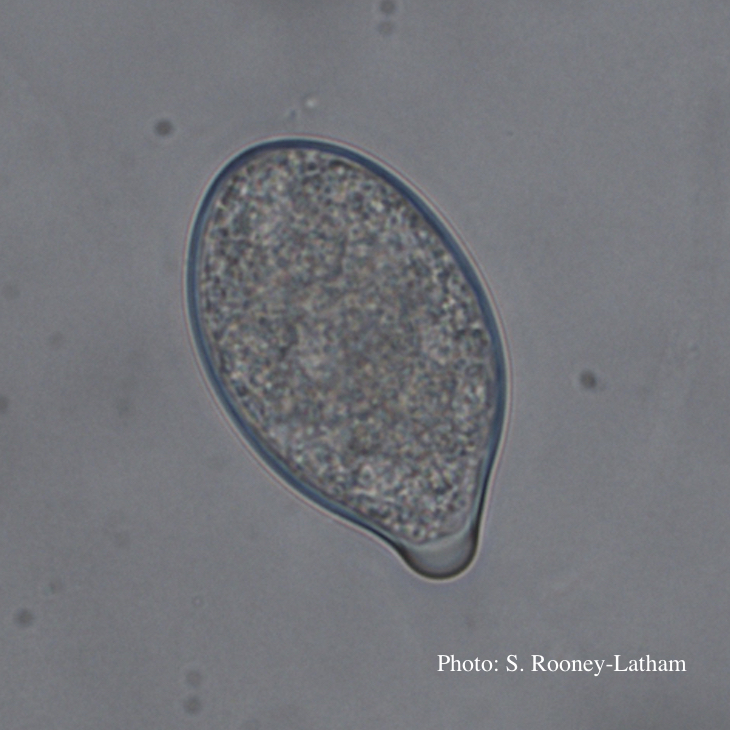 Papillate sporangium of P. tentaculata |
|
Necrotic lesion in phloem caused by P. austrocedrae  Necrotic lesion in phloem with resin pocket caused by P. austrocedrae |
P. cryptogea sporangium  Obpyriform non-papillate sporangia in water |
P. tentaculata chlamydospore  P. tentaculata chlamydospore with short hyphal projection |
|
P. siskiyouensis canker on Italian alder  Bleeding canker at the base of a tree and a sprinkler emitter (arrow) adjacent to the trunk |
P. cactorum sporangia  P. cactorum sporangia |
P. alni subsp alni sporangium 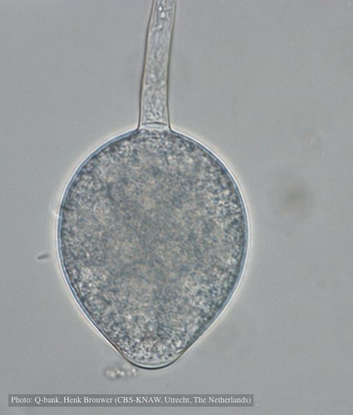 Non-papillate, non caducous sporangium, photo used with permission from Q-bank |
|
P. nemorosa colony morphology on PDA  Colony morphology on PDA at 14 days |
P. ramorum leaf symptoms on tan oak 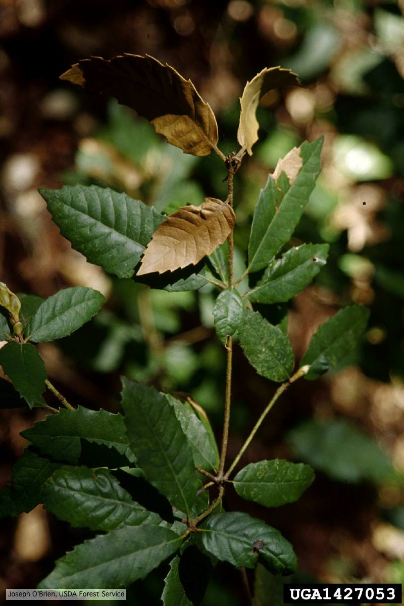 Tip symptoms on tanoak seedling (Notholithocarpus densiflorus). |
P. agathidicia oogonia 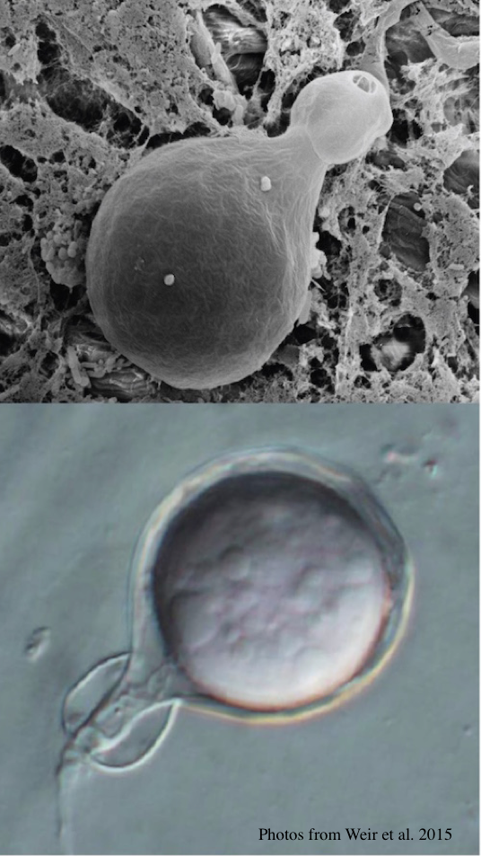 P. agathidicida oogonia with SEM (top) and light microscopy (bottom) |