

Ovoid non- papillate sporangia
Photo Gallery
Site will be retired 9/1/2026
This site is no longer being developed and will be retired on September 1, 2026. Please contact us if you have any questions or would like to provide support to continue the project.
|
P. cambivora sporangium 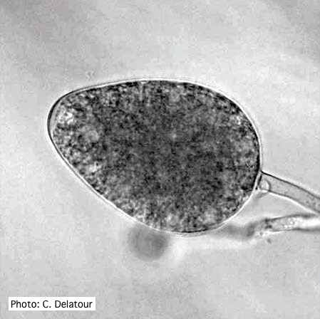 |
P. austrocedrae - Mal del ciprés, stages of decline 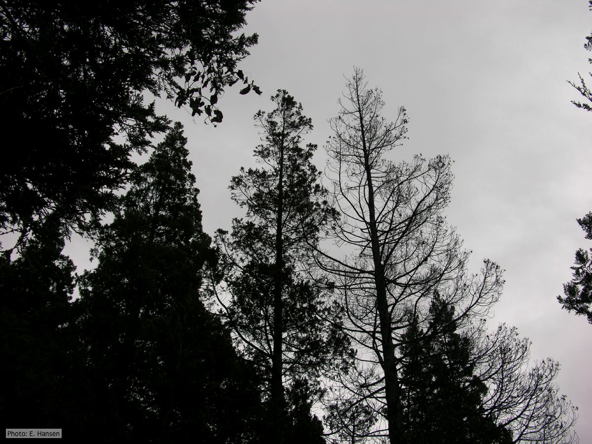 Mal del ciprés, stages of decline |
P. cinnamomi hyphal swelling 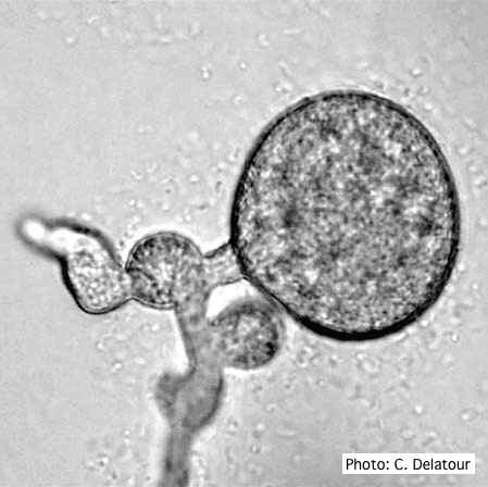 P. cinnamomi hyphal swelling (or thin walled chlamydospores) |
|
Vehicle washing  Truck washing to avoid spread of P. lateralis |
P. tentaculata disease symptoms on sticky monkey flower 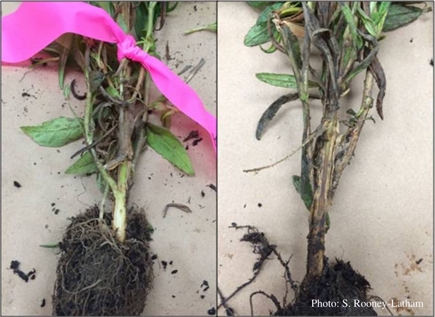 Crown and root rot (left) on sticky monkey flower (Diplacus aurantiacus) compared with a control (right) |
P. agathidicia sporangia 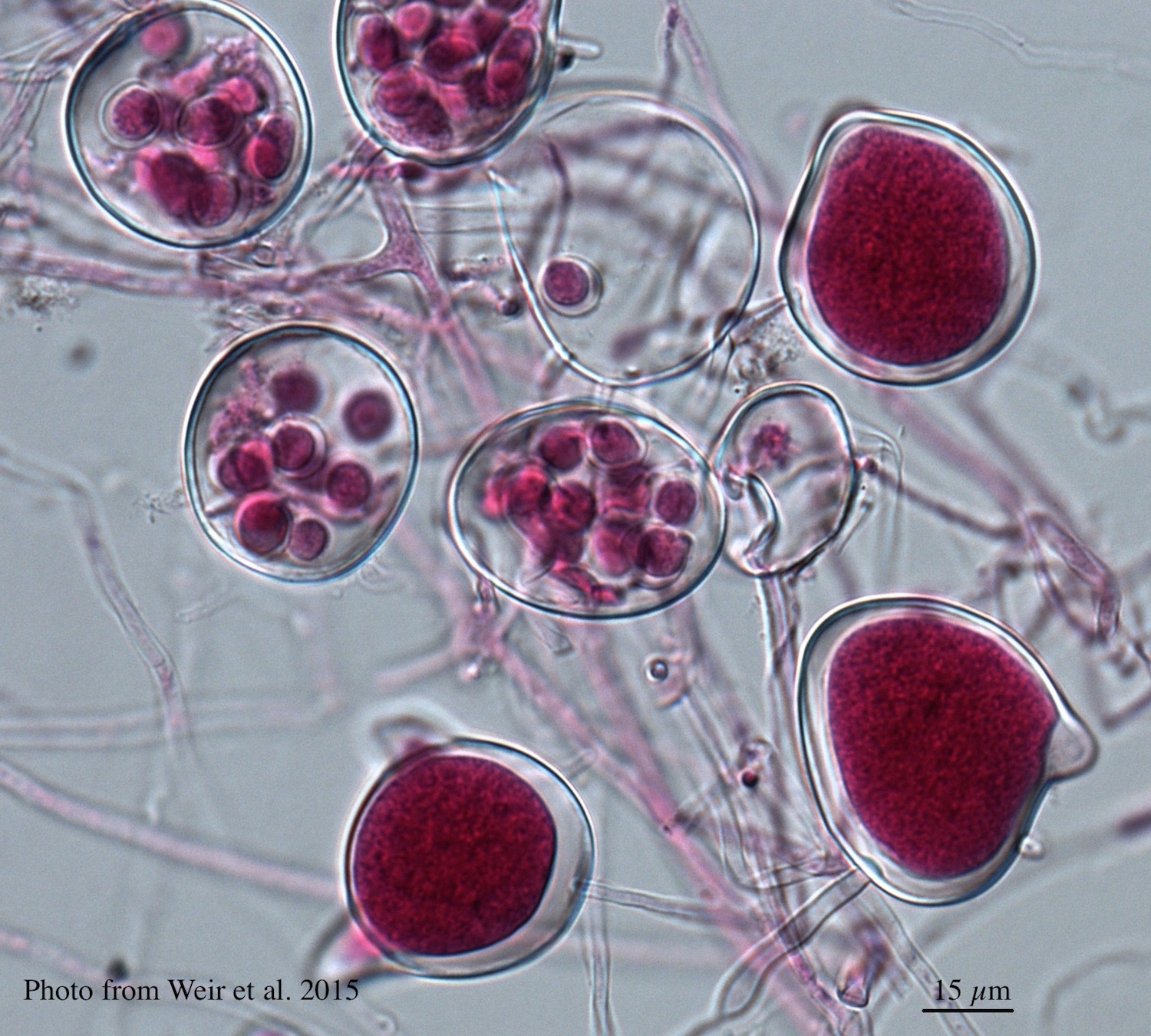 Differentiation of the cytoplasm within papillate sporangia into acid fuchsin stained zoospores |
|
P. kernoviae leaf wilt 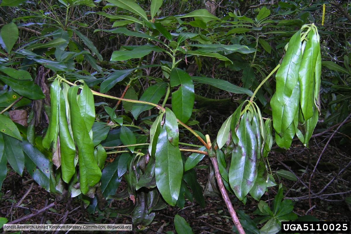 Wilted leaf of infected rhododendron |
P. cactorum colony morphology on V8  Colony morphology on V8 at 14 days |
P. alni in riparian alder, Scotland 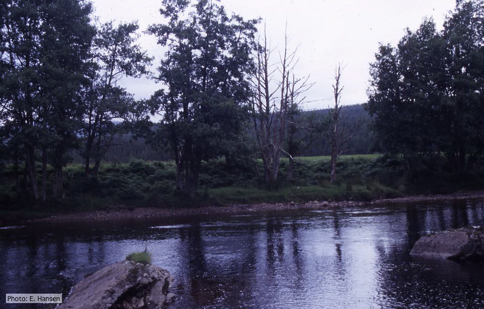 P. alni in riparian alder, Scotland |
|
P. alni oogonium 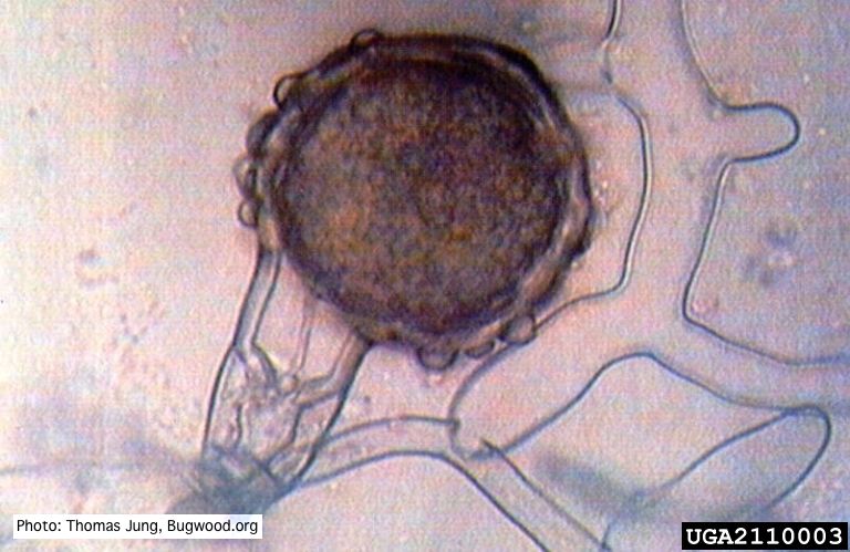 Bullate oogonium of P. alni (German variant) with oospore and amphigynous antheridium. |
P. palmivora symptoms on fruit  Brown rot on a lemon fruit caused by Phytophthora palmivora. |
P. arenaria disease symptoms on Banksia 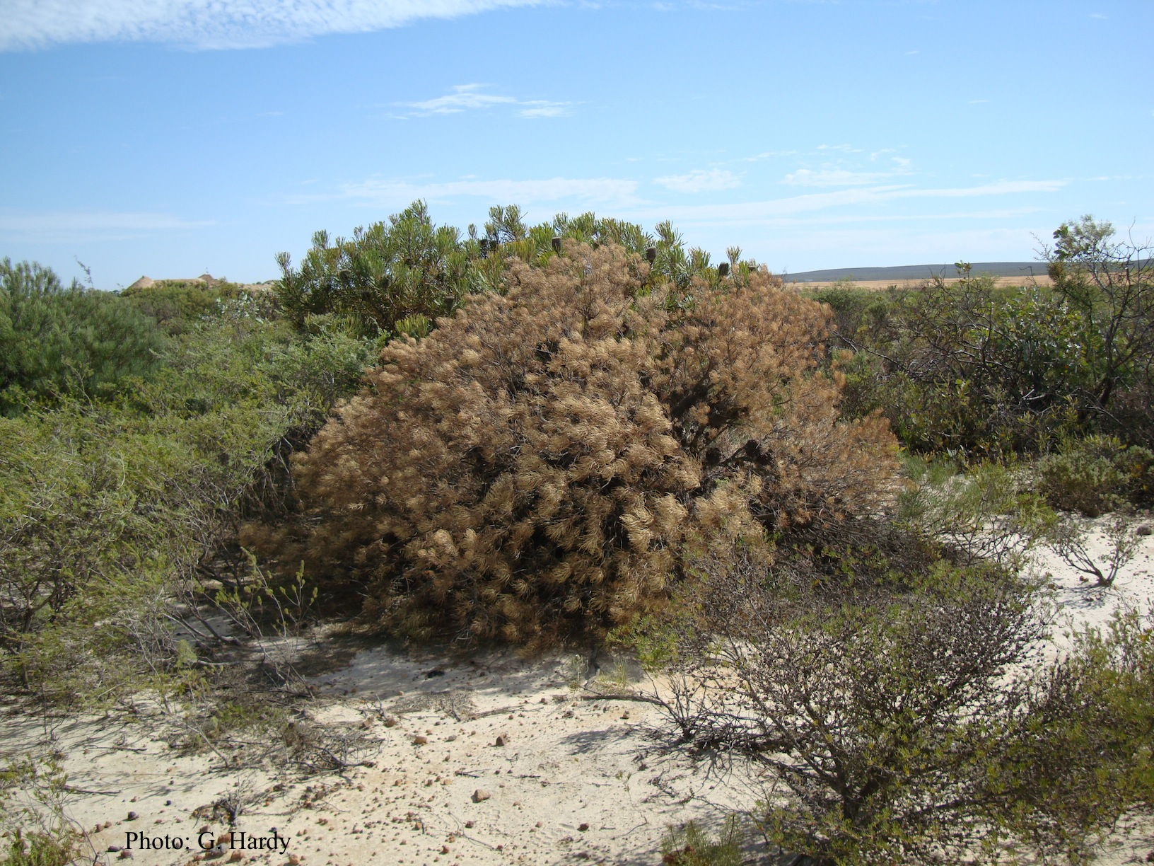 Dead Banksia sp. in a Kwongan heathland on mineral sand near Eneabba, Western Australia recently killed by root and collar rot caused by Phytophthora arenaria |