

Colony pattern after 7 days on V8 at 24C, photo from Q-bank, used with permission
Photo Gallery
Site will be retired 9/1/2026
This site is no longer being developed and will be retired on September 1, 2026. Please contact us if you have any questions or would like to provide support to continue the project.
|
P. pinifolia colony morphology on V8 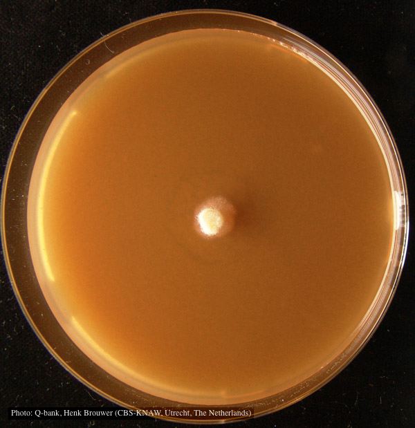 |
P. cambivora disease symptoms 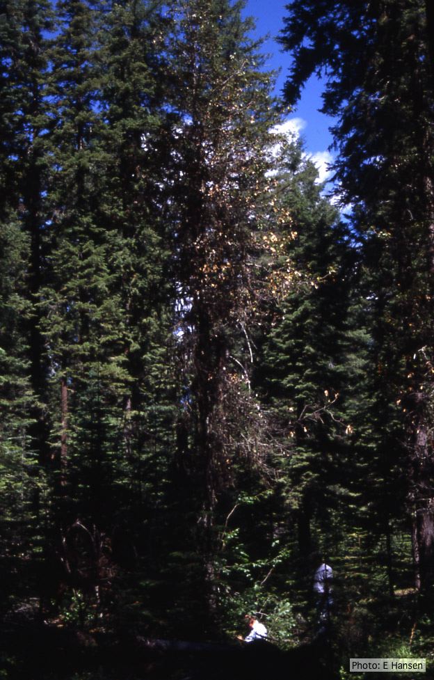 Dead and dying chinquapin infected with P. cambivora |
P. megakarya disease symptoms on Theobroma cacao fruit 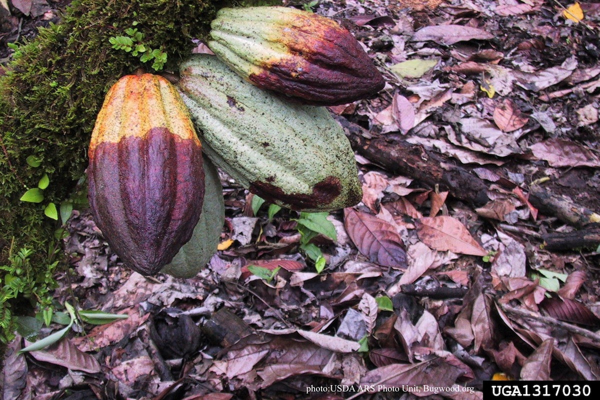 Disease symptoms on a cocoa pod |
|
Dead and healthy Port-Orford cedar trees  Chamaecyparis lawsoniana trees |
P. cinnamomi colony morphology on V8 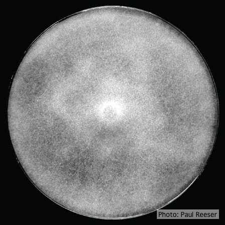 P. cinnamomi colony growth on V8 at 14 days |
Healthy Port Orford Cedar tree 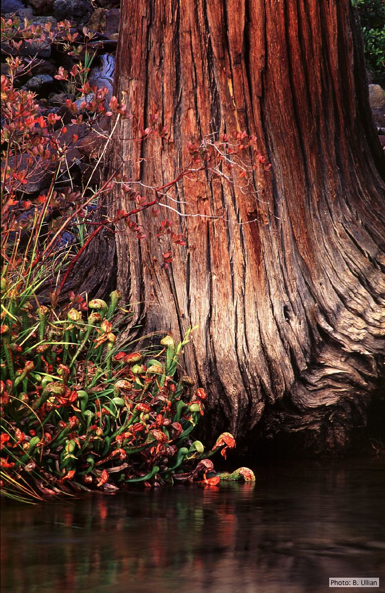 Healthy Chamaecyparis lawsoniana tree |
|
P. boehmeriae symptoms 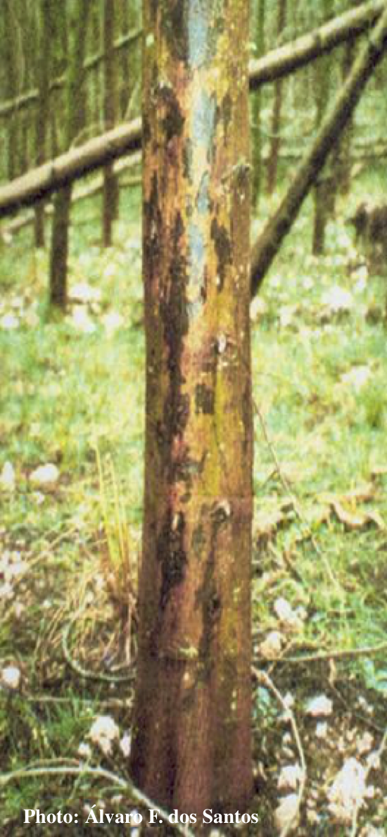 Symptoms of gummosis on black wattle (Acacia mearnsii) |
P. kernoviae canker  Bole lesion on Fagus sylvatica |
P. palmivora oogonium 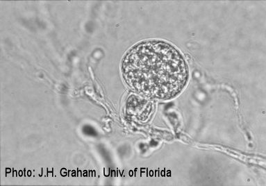 P. palmivora oogonium |
|
P. lateralis on Port Orford cedar 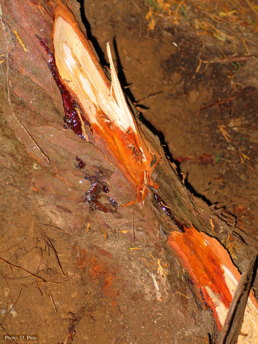 Small root lesions on Chaemacyparis lawsoniana |
P. siskiyouensis disease symptoms on Italian alder 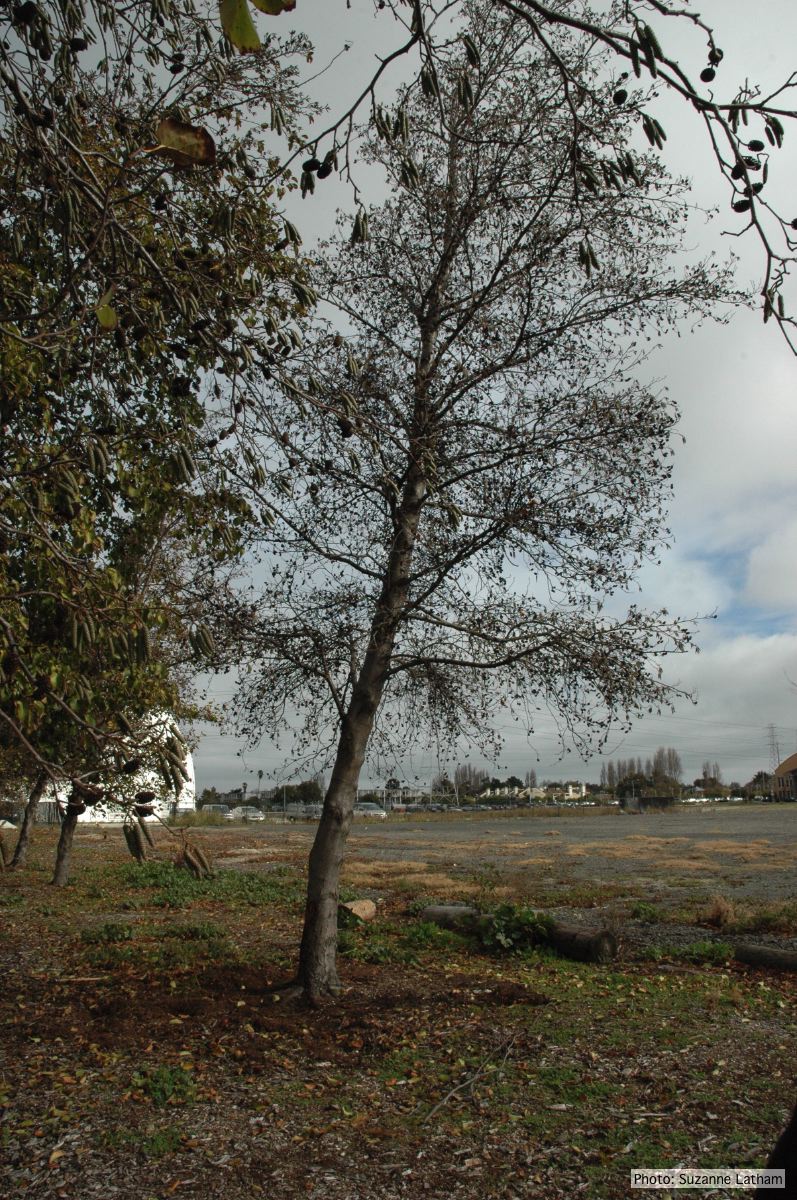 Phytophthora collar rot on Italian alder trees: standing, dead tree |
P. cambivora colony morphology on MA 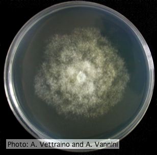 Rosacous colony morphology at 14 days at 20°C on MA |