

Death of Xanthoria and other native vegetation, Victoria, Australia
Photo Gallery
Site will be retired 9/1/2026
This site is no longer being developed and will be retired on September 1, 2026. Please contact us if you have any questions or would like to provide support to continue the project.
|
P. cinnamomi in Australia  |
P. agathadicida disease symptom 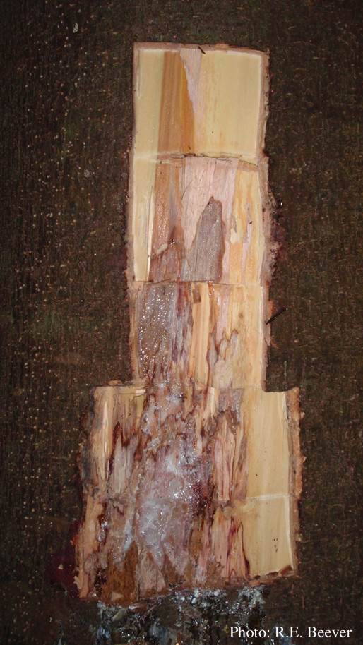 Excavated lesion, with outer bark removed showing extent of disease-front |
P. megakarya disease symptoms on Theobroma cacao

Symptoms of black pod disease of cocoa (T. cacao)
|
|
P. cinnamomi colony morphology on PDA  P. cinnamomi colony growth on PDA at 14 days |
P. cactorum bleeding canker  Bleeding canker on red oak (Quercus rubra) |
P. agathidicia growth on V8  Colony morphology of ex-holotype ICMP 17027 after 10-days incubation at 20°C in the dark |
|
P. lateralis on Port Orford cedar  Typical decline of Chaemacyparis lawsoniana in Landrévarzec, France |
P. ramorum colony morphology on CMA PARP 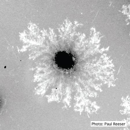 P. ramorum colony morphology on CMA PARP |
P. cinnamomi on Banksia  Death of woodland Banksia, Western Australia |
|
P. pluvialis - appearance of new growth 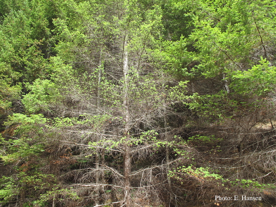 Tufted appearance of new growth from surviving buds on Douglas-fir, one year after defoliation. |
P. palmivora colony morphology on V8 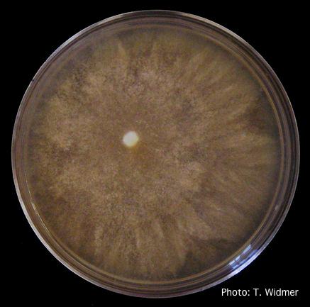 Growth of P. palmivora on V8 agar |
P. pluvialis symptoms on Douglas-fir 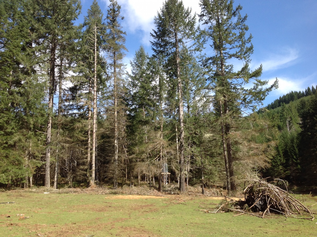 P. pluvialis symptoms of red needle cast on Douglas-fir, western Oregon 2015 |