

Asymmetrical sporangium, photo from Q-bank, used with permission
Photo Gallery
Site will be retired 9/1/2026
This site is no longer being developed and will be retired on September 1, 2026. Please contact us if you have any questions or would like to provide support to continue the project.
|
P. kernoviae sporangium 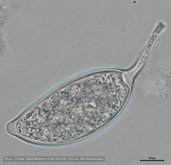 |
P. agathadicida disease symptom 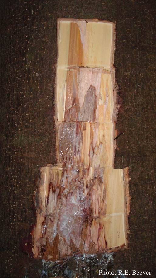 Excavated lesion, with outer bark removed showing extent of disease-front |
P. cambivora oogonium  Bullate oogonium and and two-celled amphigynous antheridium |
|
P. pinifolia hyphal growth 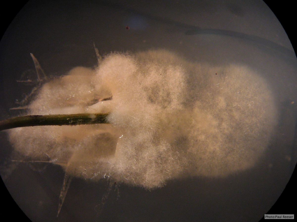 P. pinifolia pathogen growing from infected needle on selective agar |
P. austrocedrae hyphal swellings in liquid media drawing  Morphology of hyphae of Phytophthora austrocedrae, from Greslebin et al. 2007 |
P. frigida symptoms 4 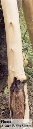 Black wattle timber with symptoms of gummosis |
|
P. alni in alder forest, Germany, with T. Jung 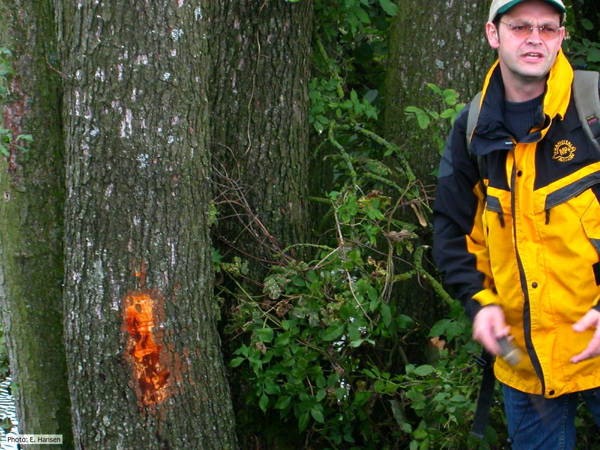 P. alni in alder forest, Germany, with T. Jung |
P. siskiyouensis colony morphology on V8 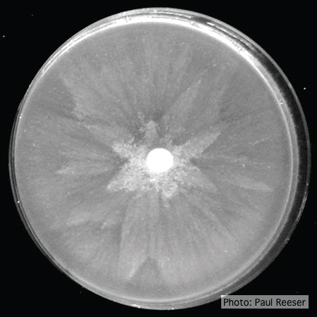 Colony morphology on V8 at 14 days |
P. austrocedrae - Mal del ciprés, stages of decline  Mal del ciprés, stages of decline |
|
P. nicotianae sporangia  P. nicotianae overview of sporangia 40x. Photo from Q-bank: www.q-bank.eu, Henk Brouwer (CBS-KNAW, Utrecht, The Netherlands) |
P. siskiyouensis bleeding canker 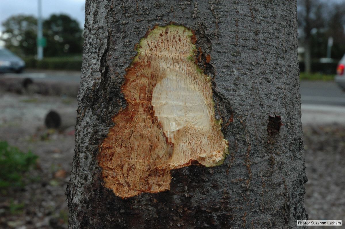 Close-up of margin area of bole lesions under the bark of a bleeding canker |
P. cryptogea colony morpholgy on PDA 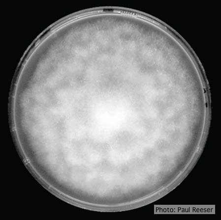 Colony morphology on PDA at 14 days |