

Sporangia (sporangiospores) showing sympodial branching
Photo Gallery
Site will be retired 9/1/2026
This site is no longer being developed and will be retired on September 1, 2026. Please contact us if you have any questions or would like to provide support to continue the project.
|
P. palmivora sporangia 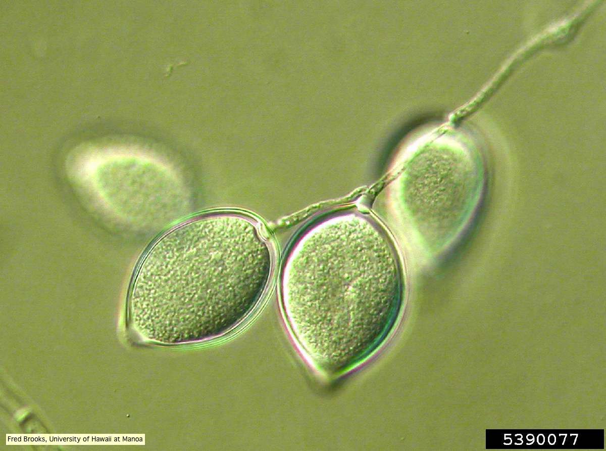 |
P. alni oogonia 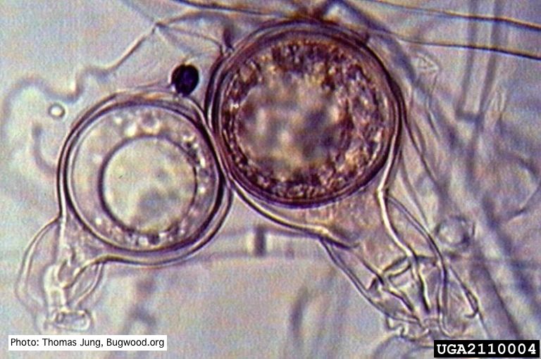 Smooth-walled oogonium of P. alni (Swedish variant) with oospore and amphigynous antheridium. |
P. cambivora symptoms  Bleeding canker, European beech, OSU campus |
|
P. cinnamomi colony morphology on PDA  P. cinnamomi colony growth on PDA at 14 days |
P. austrocedrae hyphal swellings in liquid media drawing  Morphology of hyphae of Phytophthora austrocedrae, from Greslebin et al. 2007 |
Port Orford Cedar Hedge row  Chamaecyparis lawsoniana residential hedge row with alive and dead trees |
|
P. tentaculata sporangium 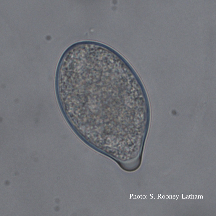 Papillate sporangium of P. tentaculata |
Chlamydospore of P. lateralis 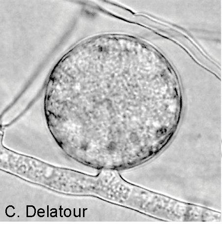 Terminal chlamydospore on a short side stalk |
P. pinifolia on Pinus radiata  Pinus radiata, note Infected needles at right angles to stem |
|
P. kernoviae leaf wilt 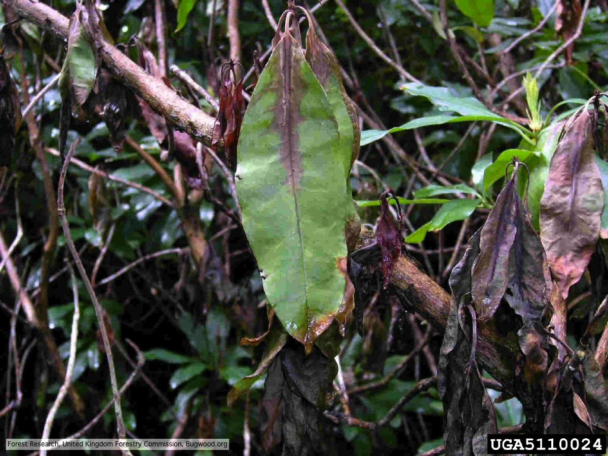 Wilted leaf of infected rhododendron |
P. ramorum sporangium  Deciduous sporangium, photo from Q-bank, used with permission |
P. lateralis on Port Orford cedar 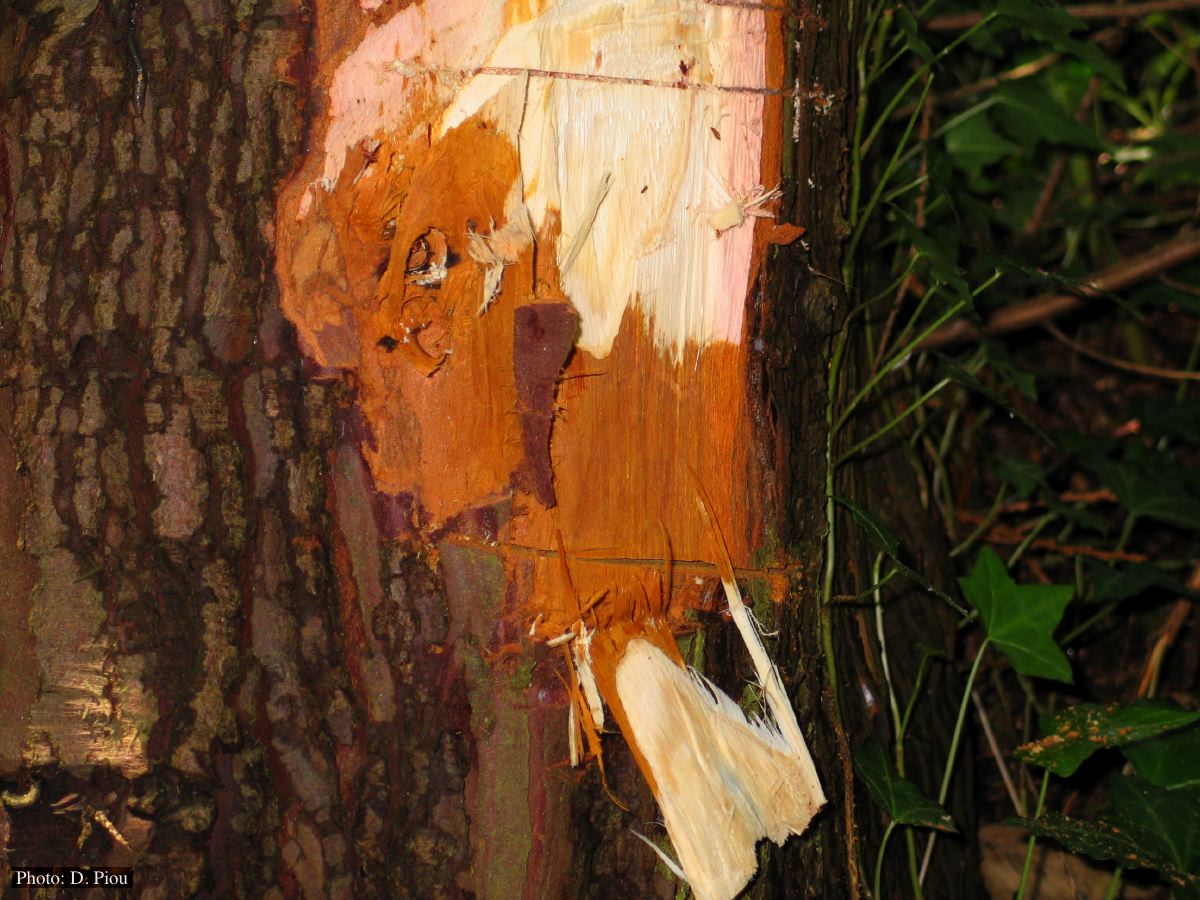 Collar lesion on Chaemacyparis lawsoniana in Landrévarzec, France |