

Oogonia with and without brown pigment, photo from Q-bank, used with permission
Photo Gallery
|
P. austrocedrae oogonium 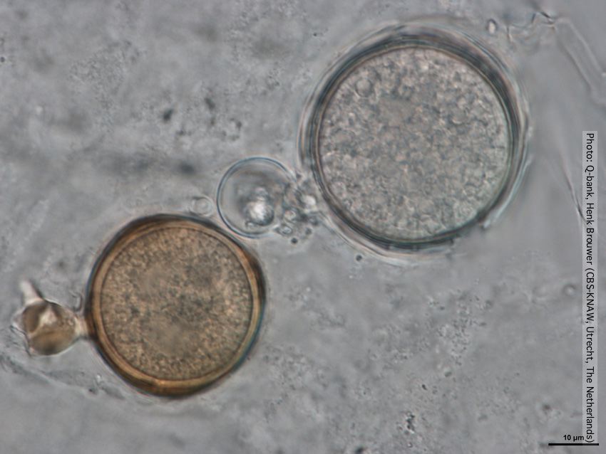 |
P. pinifolia colony morphology on CMA-NARP 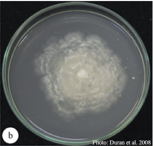 Colony morphology of P. pinifolia at 20°C on CMA-NARP after 3 weeks. From Duran et al. 2008 |
P. cambivora oogonium 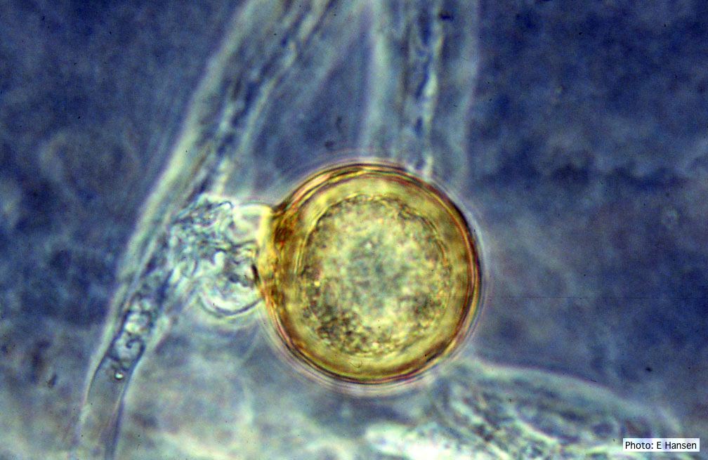 P. cambivora oogonium with antheridium |
|
P. frigida symptoms 1 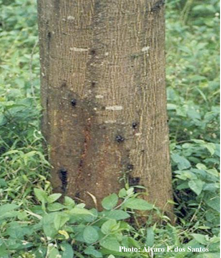 Symptoms of gummosis on black wattle |
P. agathidicia sporangia 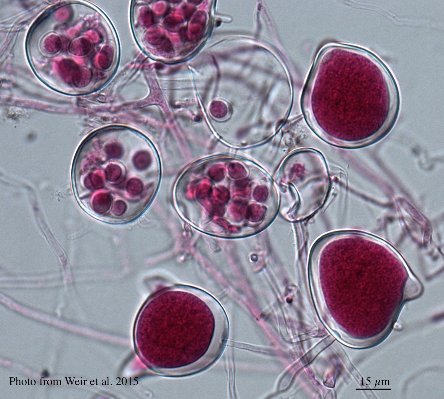 Differentiation of the cytoplasm within papillate sporangia into acid fuchsin stained zoospores |
P. agathidicida growth on MEA  Diffuse, non-patterned, colony morphology of ICMP 16471 (the original “Gadgil isolate”) after 10-days incubation at 20°C in the dark |
|
P. chlamydospora chlamydospore  Phytophthora chlamydospora chlamydospore in agar. Bar is 20µm. |
Necrotic lesion in phloem caused by P. austrocedrae  Necrotic lesion in phloem with resin pocket caused by P. austrocedrae |
P. frigida chlamydospore 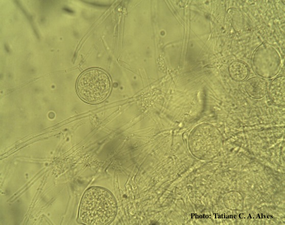 Globose chlamydospores of P. frigida |
|
P. katsurae growth morphology on β-CMA 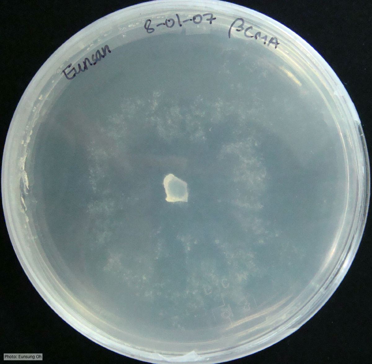 Growth morphology at 7 days on β-CMA |
Boot wash to station to control spread of P. agathidicida 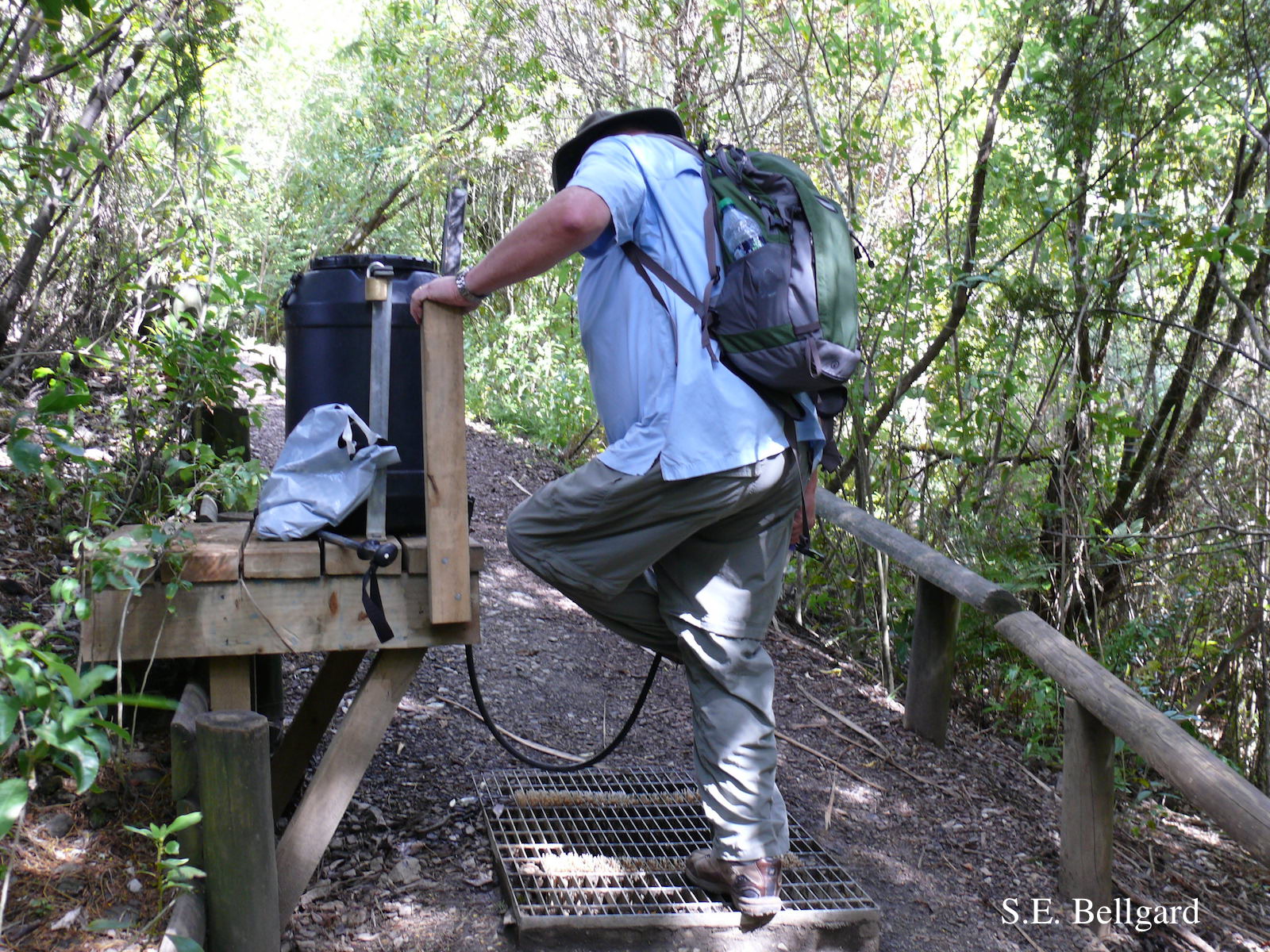 Use of hypochlorite solution applied through a “livestock drench-gun”, integrated with a soil grate to allow potentially contaminated soil to be collected |
P. boehmeriae chlamydospore 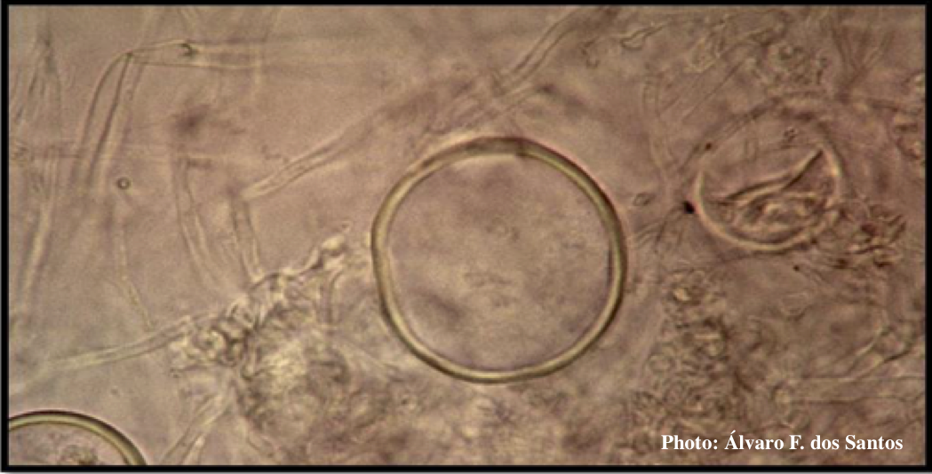 Globose chlamydospore of P. boehmeriae |