

P. ramorum colony morphology on CMA PARP
Photo Gallery
|
P. ramorum colony morphology on CMA PARP 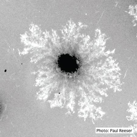 |
P. pseudosyringae sporangia  Ovoid, semipapillate sporangia showing sympodial development of sporangiophore |
P. cinnamomi on Fraser fir 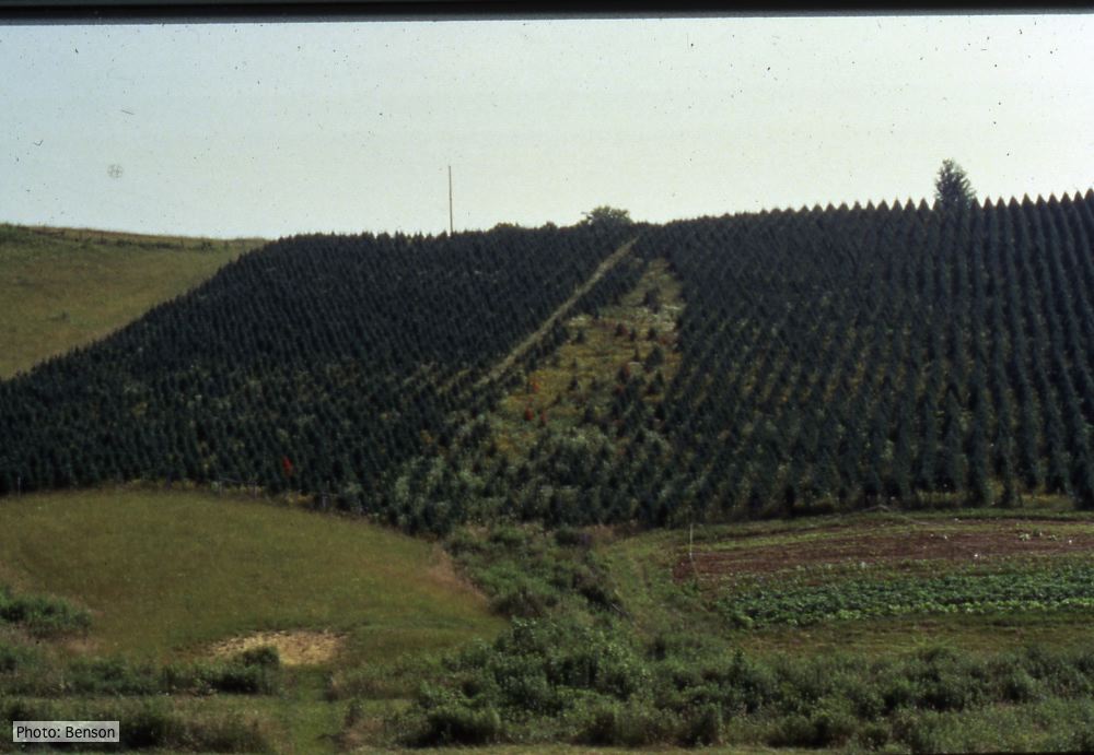 Frasier fir Christmas trees, North Carolina |
|
P. nicotianae sporangia  P. nicotianae overview of sporangia 40x. Photo from Q-bank: www.q-bank.eu, Henk Brouwer (CBS-KNAW, Utrecht, The Netherlands) |
Growth of P. megakarya on V8 agar 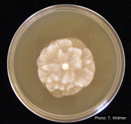 Growth of P. megakarya on V8 agar |
Comparative gametangial morphology of Phytophthora Clade 5 species  Comparative gametangial morphology of Phytophthora Clade 5 species, with SEM (top) and light microscopy (bottom). P. heveae has smooth walled oogonia with funnel-shaped, amphigynous antheridia. P. agathidicida has mildly stipulate oogonia with globose amphigynous antheridia. P.cocois has mildly bullate oogonia with reflexed amphigynous antheridia. P. castaneae has coarsely bullate oogonium with rugose protuberances and narrow amphigynous antheridia (Weir et al. 2015). |
|
P. chlamydospora sporangium 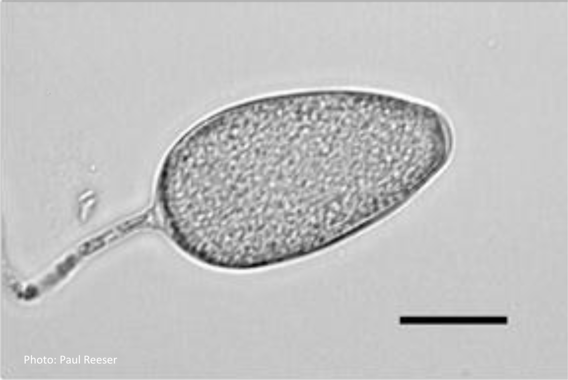 Phytophthora chlamydospora sporangium in water. Bar is 20µm. |
P. pluvialis oogonium and antheridium 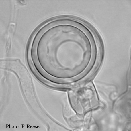 Oogonium and oospore with amphigynous antheridium |
P. pluvialis on Pinus radiata in New Zealand  Typical red needle cast symptoms along a twig. Lesions begin at the base of the needle which subsequently turns brown and is cast from the twig. |
|
P. cryptogea hyphal swellings 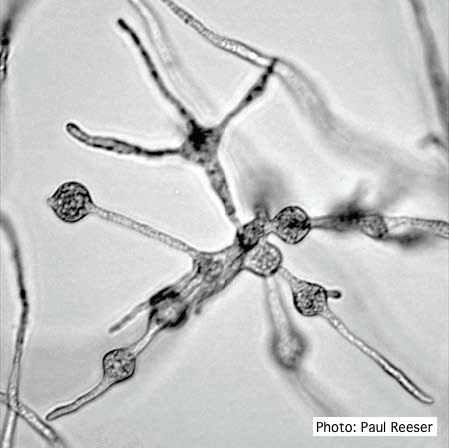 Cluster of small, angular to globose hyphal swellings formed in water |
P. tentaculata on V-8 media 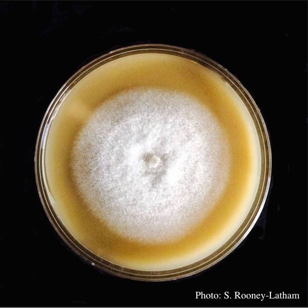 Culture of P. tentaculata on V-8 media |
P. cactorum sporangia 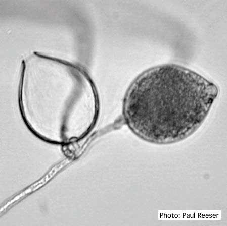 Broadly ovoid, papillate sporangia in water. |