

P. pluvialis hyphal swellings on agar
Photo Gallery
|
P. pluvialis hyphal swellings 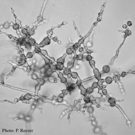 |
P. kernoviae colony morphology on CMA PARPH 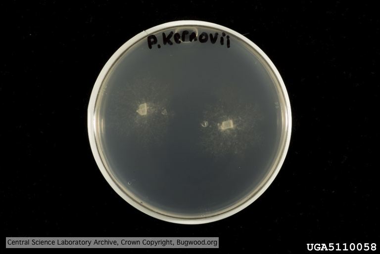 Organism grown on CMA PARP[H]; Plant disease 70, 1038-1043 |
P. chlamydospora chlamydospore 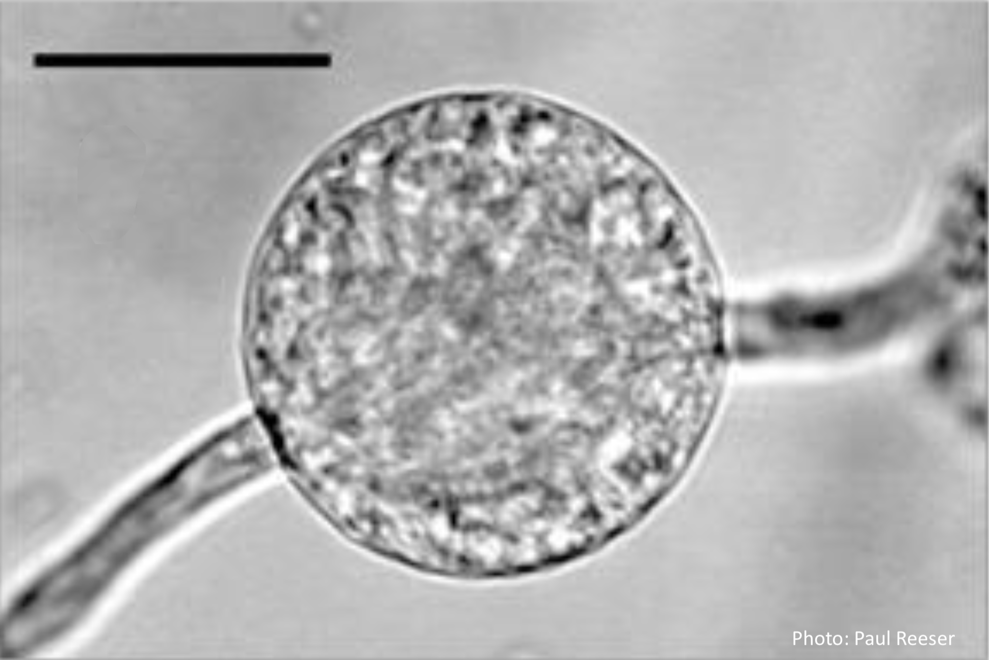 Phytophthora chlamydospora chlamydospore in agar. Bar is 20µm. |
|
P. pluvialis symptoms on Douglas-fir 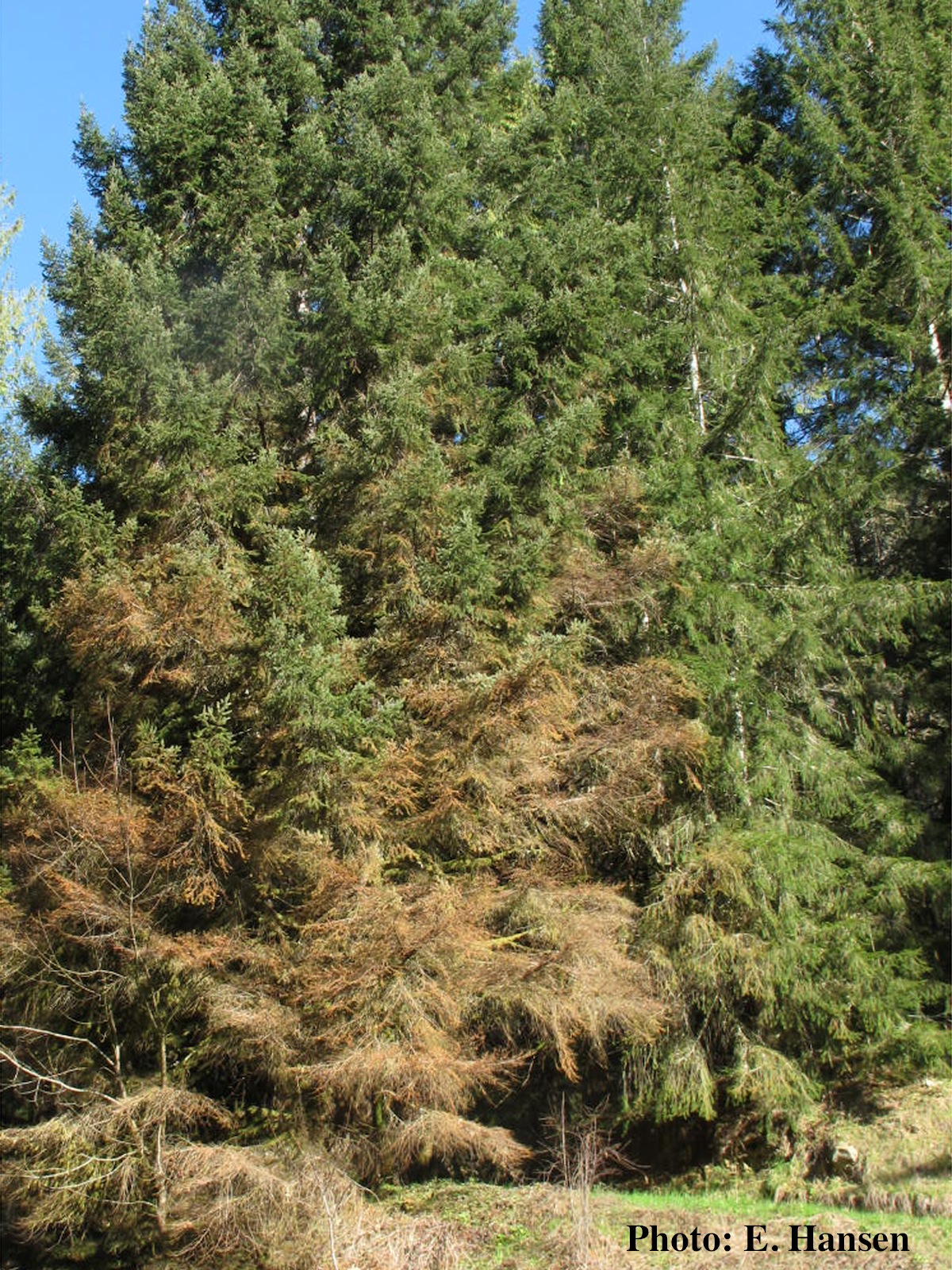 Red needle cast symptoms on Douglas-fir in western Oregon, 2015 |
P. chlamydospora colony morphology on carrot agar 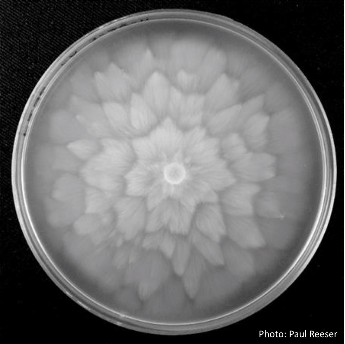 P. chlamydospora colony morphology on carrot agar |
P. siskiyouensis bleeding canker  Bole lesions in the tissues under the bark of a bleeding canker: discoloration in the secondary phloem tissue |
|
P. austrocedrae - sporangia drawings 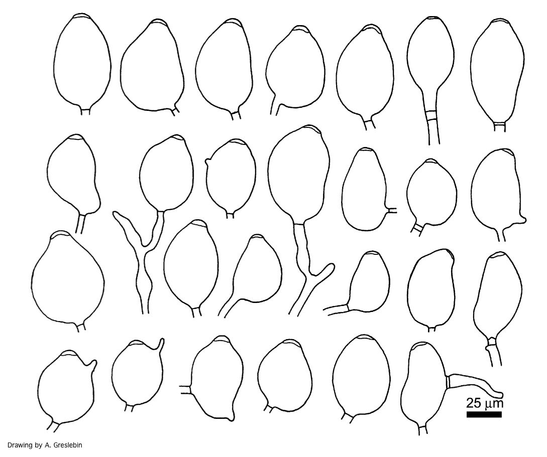 Phytophthora austrocedrae. Morphology of sporangia. Bar: 25 mm. Greslebin et al. 2007 |
P. agathidicida growth on CMA 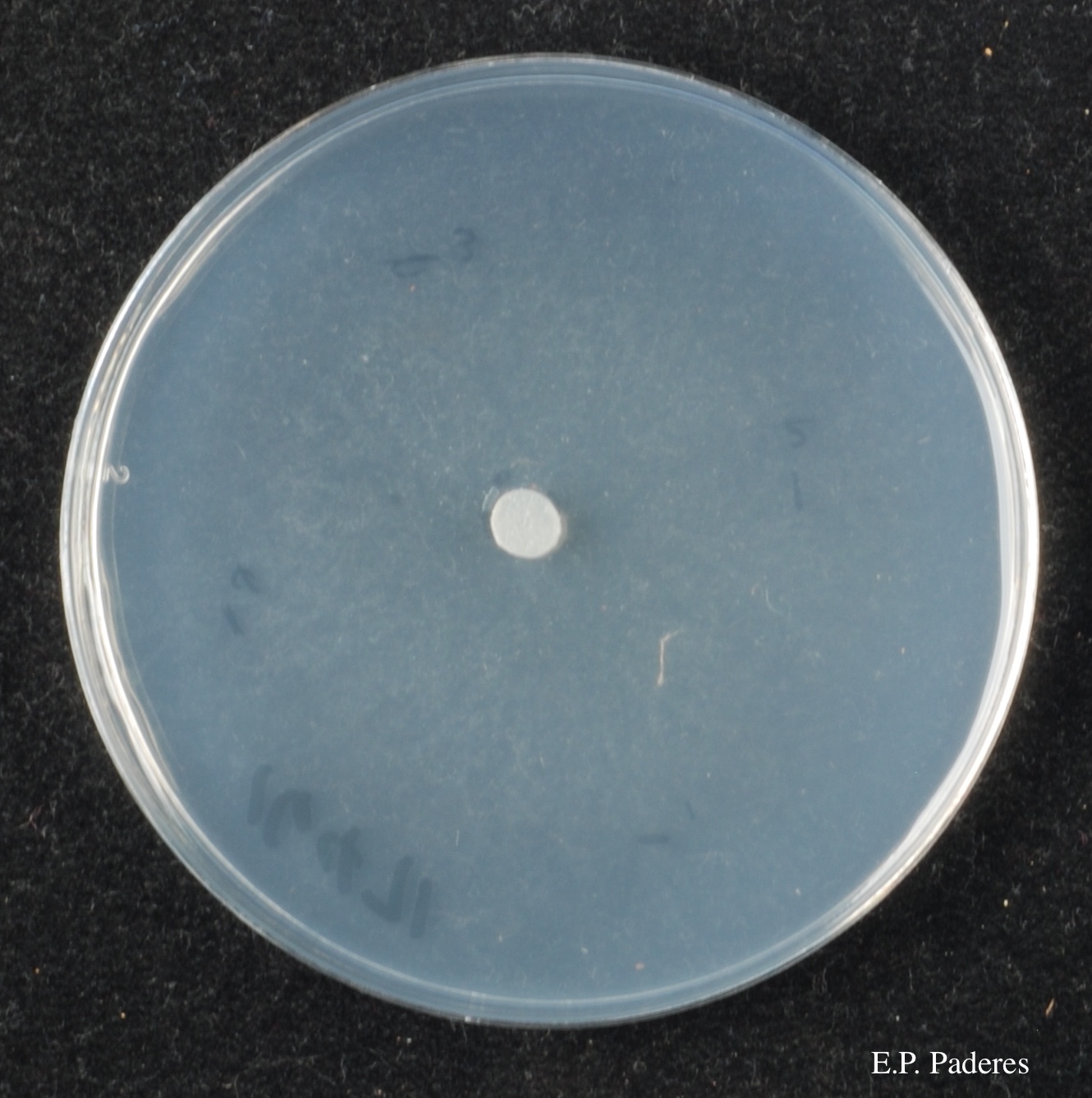 Diffuse, non-patterned, colony morphology of ICMP 16471 (the original “Gadgil isolate”) after 10-days incubation at 20°C in the dark |
P. cinnamomi on Jarrah 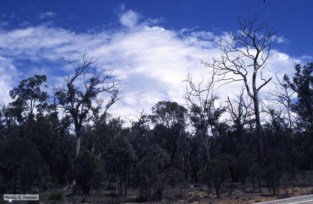 Dieback in Jarrah, Western Australia |
|
P. cinnamomi sporangium 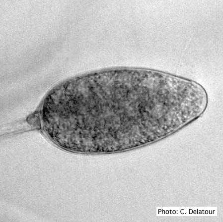 P. cinnamomi sporangium |
P. chlamydospora sporangium 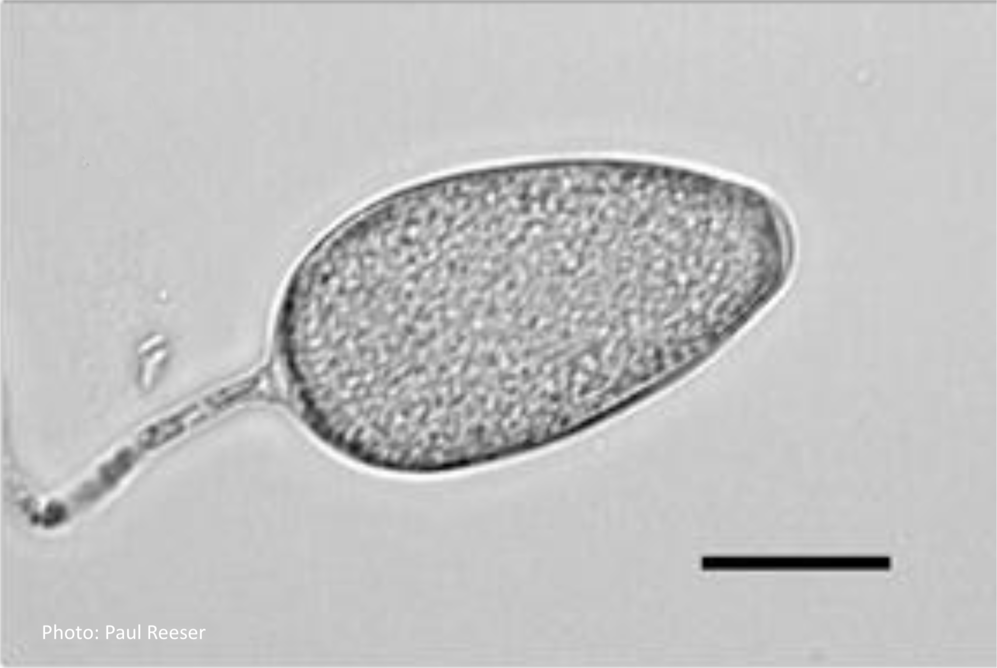 Phytophthora chlamydospora sporangium in water. Bar is 20µm. |
P. cactorum bleeding canker 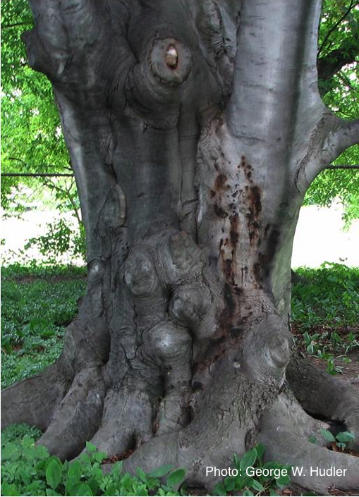 Bleeding canker on European beech (Fagus sylvatica) |