

Phytophthora chlamydospora sporangia in water, showing internal proliferation. Bar is 20 µm.
Photo Gallery
Site will be retired 9/1/2026
This site is no longer being developed and will be retired on September 1, 2026. Please contact us if you have any questions or would like to provide support to continue the project.
|
Phytophthora chlamydospora sporangium 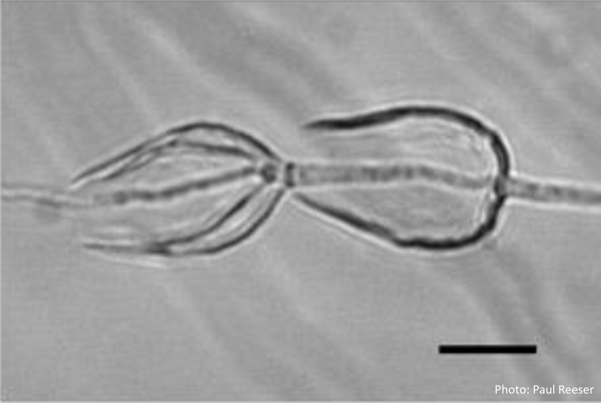 |
P. chlamydospora sporangium 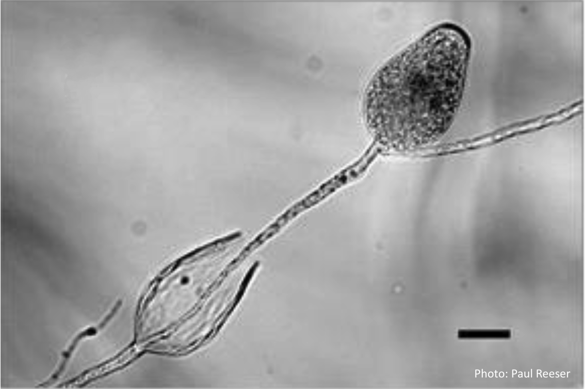 Phytophthora chlamydospora sporangia in water, showing subsporangial elongation. Bar is 20 µm. |
P. chlamydospora colony morphology on V8 agar 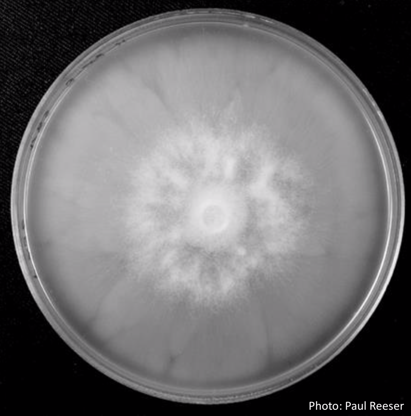 P. chlamydospora colony morphology on V8 agar |
|
P. chlamydospora colony morphology on carrot agar 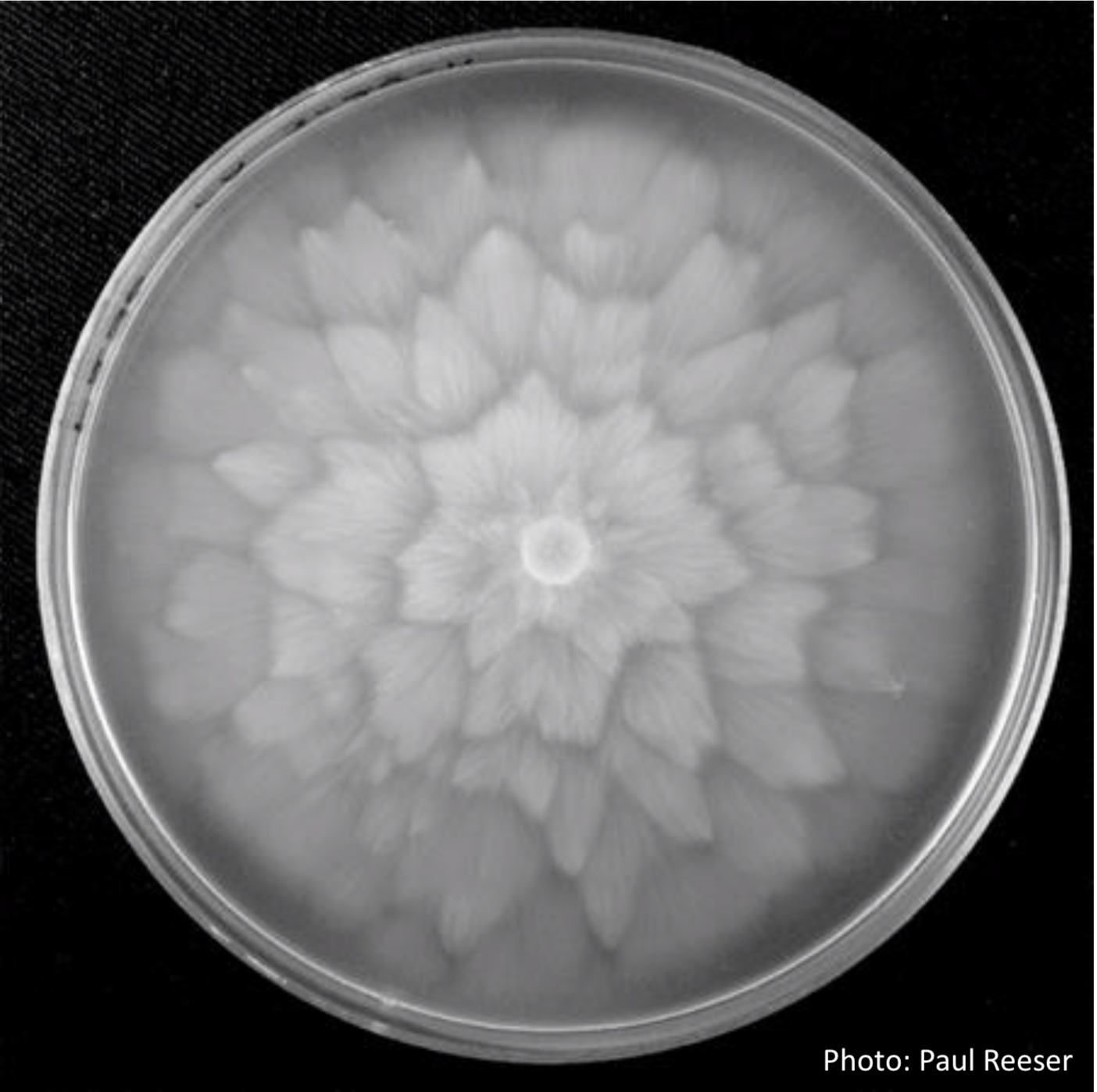 P. chlamydospora colony morphology on carrot agar |
P. chlamydospora sporangium  Phytophthora chlamydospora sporangium in water. Bar is 20µm. |
P. chlamydospora sporangium 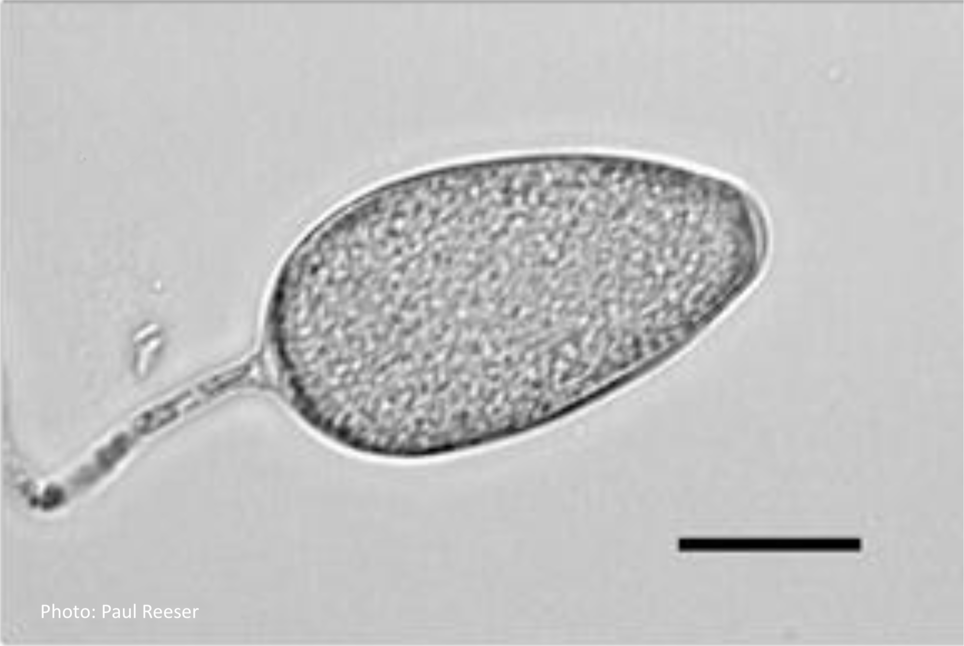 Phytophthora chlamydospora sporangium in water. Bar is 20µm. |
|
P. chlamydospora chlamydospore 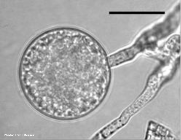 Phytophthora chlamydospora chlamydospore in agar. Bar is 20 µm. |
P. chlamydospora chlamydospore  Phytophthora chlamydospora chlamydospore in agar. Bar is 20µm. |
P. chlamydospora hyphal swellings 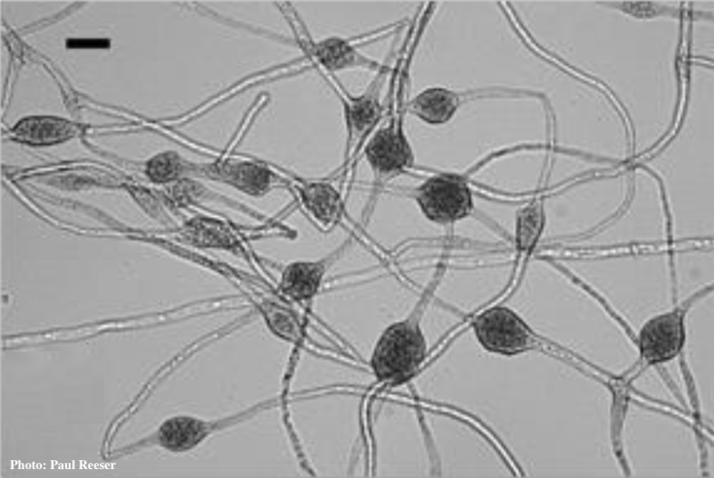 Phytophthora chlamydospora chlamydospore in agar. Bar is 20µm.
|
|
P. chlamydospora chlamydospore 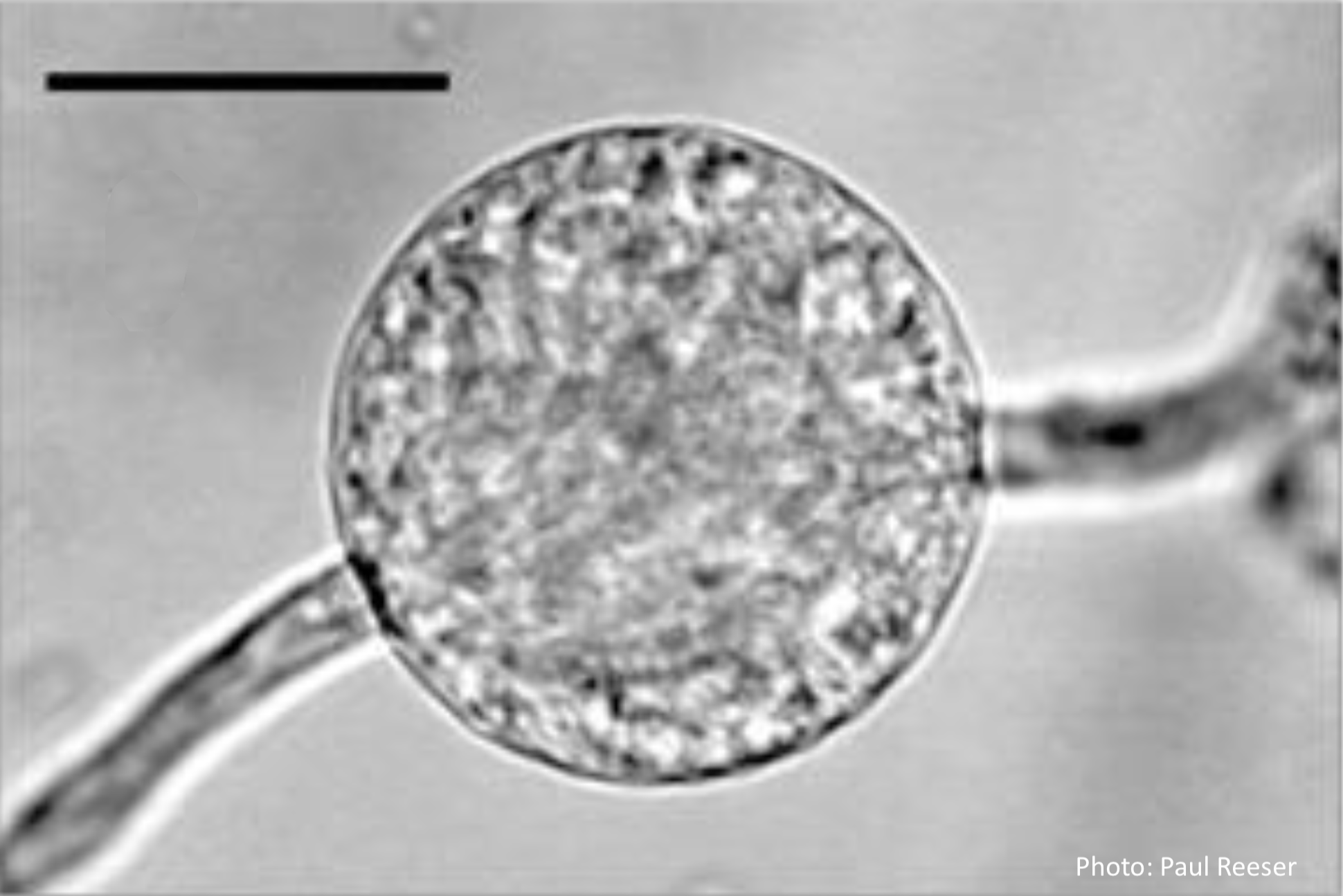 Phytophthora chlamydospora chlamydospore in agar. Bar is 20µm. |